Premature atrial and ventricular contractions !
Extra heartbeats that come too early, felt as a skip or a thump. Very common and usually harmless, but sometimes a sign of heart disease.
Updated 2026-11-01
Structures involved
Tap one to see what it is and open it in 3D.
- Right atrium
- Left ventricle
- Sinoatrial (SA) node
- Left atrium
- Right ventricle
- Atrioventricular (AV) node
- Bundle of His
- Bundle branches
- Pulmonary veins
- Pulmonary trunk
Why it happens
- How arrhythmias arise
- The SA node pacemaker potential
- The ECG in potassium and calcium disorders
- Na⁺/K⁺-ATPase (sodium pump)
What it is
Premature contractions (extrasystoles) are beats that arise earlier than the next expected sinus beat from an ectopic focus outside the sinoatrial node. Premature atrial contractions (PACs) start in the atria and premature ventricular contractions (PVCs) in the ventricles. They are the most common arrhythmias. Isolated beats in a normal heart are usually benign, but a high burden can cause symptoms or cardiomyopathy, and they may reflect structural heart disease or a metabolic problem.
Who gets it
Very common: some PACs and PVCs appear on 24-hour monitoring in most adults, and their frequency rises with age. They are seen more often in people with hypertension, heart failure, ischaemic or Chagas heart disease. In Colombia and Latin America, frequent or complex PVCs in someone from a rural endemic area should prompt testing for Chagas disease.
Causes
- Idiopathic in a normal heart (for example from the right ventricular outflow tract)
- Stimulants: caffeine, nicotine, alcohol, cocaine and amphetamines, some decongestants
- Electrolyte disorders: low potassium or magnesium
- Structural heart disease: ischaemic heart disease, cardiomyopathies (including Chagas), valve disease, heart failure
- Hyperthyroidism, hypoxia, sleep apnoea, stress and anxiety, digoxin toxicity
Risk factors
- Older age
- Hypertension and structural heart disease
- Caffeine, alcohol, tobacco and stimulant use
- Sleep deprivation and emotional stress
- Electrolyte disturbances, often from diuretics
Pathophysiology
Three mechanisms produce premature beats: enhanced automaticity of an ectopic focus, triggered activity from afterdepolarisations (as in digoxin toxicity or hypokalaemia), and micro re-entry around scar. A PAC is conducted through the AV node, giving a narrow QRS preceded by an abnormal P wave, and often resets the sinus node (non-compensatory pause). A PVC spreads slowly through ventricular muscle rather than the conduction system, giving a wide, bizarre QRS without a preceding P wave, usually followed by a full compensatory pause. The beat after the pause is stronger, which patients feel as a thump. A PVC burden above roughly 10 to 15 % of beats can cause dyssynchrony and a reversible PVC-induced cardiomyopathy.
Classification
- By origin: atrial (PAC), junctional, ventricular (PVC)
- PVC morphology: unifocal (monomorphic) versus multifocal (polymorphic)
- Patterns: bigeminy (every other beat), trigeminy, couplets; three or more in a row is non-sustained ventricular tachycardia
- R-on-T PVCs (falling on the T wave), which can trigger ventricular fibrillation in an ischaemic heart
Symptoms and signs
- Many people have no symptoms
- Sensation of a skipped beat, a pause or a strong thump in the chest
- Fluttering or palpitations, often noticed at rest or lying down
- Light-headedness with frequent beats
- Fatigue or dyspnoea when the burden is high or heart function is reduced
Diagnosis
- Clinical
- History of palpitations, triggers (caffeine, alcohol, stimulants, sleep), drugs and family history; examination for murmurs and heart failure. An irregular pulse with occasional pauses suggests premature beats. The aim is to separate benign isolated ectopy from beats that signal heart disease.
- Tests
- 12-lead ECG identifies the type and morphology. Holter or longer ambulatory monitoring measures the burden and detects runs. Potassium, magnesium, thyroid function and, where relevant, digoxin level; Chagas serology in people from endemic areas. Exercise testing: PVCs that disappear with exercise are usually benign, while those that increase deserve further study.
- Imaging
- Echocardiography to look for structural heart disease and PVC-induced cardiomyopathy when PVCs are frequent, complex or symptomatic. Cardiac magnetic resonance when echocardiography suggests cardiomyopathy.
Differential diagnosis
- Atrial fibrillation (irregularly irregular rhythm)
- Sinus arrhythmia
- Second-degree AV block (dropped beats)
- Supraventricular tachycardia
- Non-sustained ventricular tachycardia
- Anxiety or panic disorder with sinus tachycardia
Goal of treatment
Reassure when benign, remove triggers, correct the cause and treat symptoms or a high burden that threatens ventricular function.
Treatment
- Conservative
- Reassurance and explanation for isolated beats in a normal heart; no treatment needed when asymptomatic.
- Lifestyle
- Reduce or avoid caffeine, alcohol, tobacco, stimulants and decongestants; improve sleep and manage stress.
- Medical
- Correct low potassium or magnesium, treat hyperthyroidism, review drugs such as digoxin, and treat underlying heart disease.
- Medical
- Symptomatic or frequent beats: beta-blockers or non-dihydropyridine calcium channel blockers first; antiarrhythmic drugs in selected cases.
- Procedure
- Catheter ablation of the ectopic focus for frequent symptomatic PVCs, failure of drugs or PVC-induced cardiomyopathy.
- Follow-up
- Nursing: assess the patient, not just the monitor; report new, frequent, multifocal or R-on-T PVCs and runs, check potassium and magnesium, and monitor closely after myocardial infarction.
Complications
- Usually none in a normal heart
- PVC-induced cardiomyopathy with a high burden
- Triggering of atrial fibrillation (frequent PACs, especially from the pulmonary veins)
- Ventricular tachycardia or fibrillation in an ischaemic or diseased heart
- Anxiety and reduced quality of life from palpitations
Prognosis
Isolated PACs and PVCs in a structurally normal heart carry an excellent prognosis. Frequent PACs predict later atrial fibrillation, and frequent or complex PVCs with structural heart disease are linked to higher risk of arrhythmic death. PVC-induced cardiomyopathy often improves after the burden is reduced.
Prevention
- Moderating caffeine and alcohol and avoiding tobacco and stimulants
- Adequate sleep and stress management
- Control of hypertension and monitoring of electrolytes in patients on diuretics
Test yourself
Think of the answer, then tap the question to see it.
How does a PVC look on the ECG?
A wide, bizarre QRS without a preceding P wave, usually followed by a full compensatory pause.
How does a PAC look on the ECG?
An early, abnormal P wave followed by a narrow QRS, usually with a non-compensatory pause.
What is ventricular bigeminy?
A PVC after every normal sinus beat.
Which electrolytes should be checked in a patient with frequent PVCs?
Potassium and magnesium.
What can a very high PVC burden cause?
A reversible PVC-induced cardiomyopathy.
Related conditions
- Atrial fibrillation
- Atrial flutter
- Supraventricular tachycardia (AVNRT, AVRT)
- Ventricular tachycardia
- Dilated cardiomyopathy
- Heart failure
- Myocardial Infarction (Heart Attack)
- Graves disease (hyperthyroidism)
- Hypomagnesaemia
- Digoxin toxicity
- Obstructive sleep apnoea
- Panic disorder
- Arrhythmogenic right ventricular cardiomyopathy
- Cocaine and sympathomimetic toxicity
Educational information, not medical advice. For any symptom, see a health professional.
Sources: Arrhythmias - Types | NHLBI, NIH (National Heart, Lung, and Blood Institute (NHLBI), NIH, 2026); Arrhythmias: MedlinePlus Medical Encyclopedia (MedlinePlus, US National Library of Medicine, 2026).
Spotted a mistake?We are always improving for the community. Tell us what is wrong and our team will review it.Report a mistake
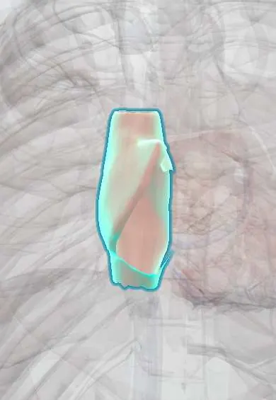
Representative model, for educational purposes only.