Conditions · Infectious and tropical diseases
Echinococcosis (hydatid disease) !
A tapeworm infection caught from dog or fox faeces that grows slow fluid-filled cysts, mostly in the liver and lungs.
Updated 2026-11-01
Structures involved
Tap one to see what it is and open it in 3D.
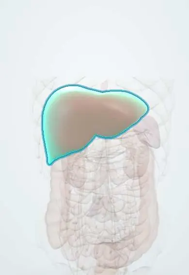
- Liver
- Right lower lobe
- Left lower lobe
- Liver segment VII
- Liver segment VIII
- Common bile duct
- Common hepatic duct
- Hepatic veins
- Inferior vena cava
- Diaphragm
- Peritoneum
- Spleen
- Pleura
- Main bronchi
- Lobar bronchi
Microorganisms that cause it
Why it happens
What it is
A zoonotic infection by the larval stage of Echinococcus tapeworms. Cystic echinococcosis (hydatid disease, E. granulosus) forms one or more slowly enlarging fluid-filled cysts, mainly in the liver (about two thirds) and lungs. Alveolar echinococcosis (E. multilocularis) forms an infiltrating, tumour-like liver mass that can invade neighbouring organs and spread at a distance.
Who gets it
Cystic echinococcosis occurs worldwide wherever dogs are fed raw offal from slaughtered livestock; in South America it is highly endemic in the sheep-raising regions of Peru, Chile, Argentina, Uruguay and southern Brazil. Colombia reports few, sporadic cases, while the neotropical polycystic form caused by E. vogeli (bush dog and paca cycle) has been described in rural Colombia and the Amazon basin. Alveolar echinococcosis is a Northern Hemisphere disease (China, Central Asia, Europe, North America) and is rare but often fatal. Worldwide more than a million people are affected at any time.
Causes
- Echinococcus granulosus (sensu lato): cystic echinococcosis; dog is the definitive host, sheep, cattle, goats and pigs are intermediate hosts
- Echinococcus multilocularis: alveolar echinococcosis; foxes and other wild canids are definitive hosts, small rodents are intermediate hosts
- Echinococcus vogeli and E. oligarthra: neotropical polycystic and unicystic forms in Central and South America
- Humans are accidental dead-end hosts infected by swallowing eggs from faeces of the definitive host
Risk factors
- Close contact with dogs that eat raw viscera of slaughtered animals
- Rural sheep or cattle farming and home slaughter
- Drinking untreated water or eating unwashed vegetables contaminated with dog faeces
- Poor hand hygiene, especially in children who play with dogs
- Hunting, trapping or handling foxes (alveolar form)
Pathophysiology
Swallowed eggs hatch in the small intestine; the released embryo (oncosphere) crosses the gut wall and travels in the portal blood, so the liver is the first filter and the lungs the second. In cystic disease the larva becomes a cyst (hydatid) with an outer host-made fibrous pericyst, a laminated acellular layer and an inner germinal layer that buds daughter cysts and protoscolices ('hydatid sand'). Growth is slow, about 1–5 cm per year, and symptoms come from mass effect, rupture into the bile ducts, bronchi or peritoneum, or secondary infection. Leakage of the antigen-rich fluid can trigger anaphylaxis, and spilled protoscolices seed new cysts. In alveolar disease the larva proliferates by outward budding like a malignant tumour, with necrosis, fibrosis and calcification, invading the hilum, vessels and diaphragm and metastasising to lung and brain.
Classification
- By species: cystic (E. granulosus), alveolar (E. multilocularis), polycystic (E. vogeli) and unicystic (E. oligarthra)
- WHO ultrasound staging of cystic liver cysts: CL (unclear), CE1 and CE2 active, CE3a/CE3b transitional, CE4 and CE5 inactive and calcified
- Alveolar echinococcosis PNM staging: extent in the liver (P), neighbouring organs (N) and metastases (M)
Symptoms and signs
- Often silent for years and found by chance on ultrasound or chest X-ray
- Liver cyst: right upper quadrant fullness or pain, palpable mass or hepatomegaly, nausea
- Jaundice, colicky pain and fever when the cyst ruptures into the biliary tree
- Lung cyst: cough, chest pain, haemoptysis or dyspnoea
- Coughing up salty clear fluid or membrane fragments after rupture into a bronchus (vomica)
- Alveolar form: weight loss, jaundice and a hard irregular liver, mimicking liver cancer
Diagnosis
- Clinical
- Suspected in a person from a livestock-raising area, with dog contact, who has a cystic mass in the liver or lung. Eosinophilia is inconstant and appears mainly with leakage. Diagnostic puncture is avoided unless done under drug cover as part of a planned procedure, because of the risk of anaphylaxis and spread.
- Tests
- Serology (ELISA or indirect haemagglutination, confirmed by immunoblot) supports the diagnosis; it is more often positive in liver than in lung cysts, and a negative test does not exclude disease. Liver function tests if biliary involvement is suspected. Microscopy of fluid or membranes removed at surgery or PAIR shows protoscolices and hooklets.
- Imaging
- Ultrasound is the key test and allows WHO staging: a cyst with a double wall, floating membranes ('water-lily sign'), daughter cysts ('honeycomb') or calcification. CT or MRI defines extent and relation to bile ducts and vessels; chest X-ray shows round lung opacities, with the air-crescent or water-lily sign after rupture. In the alveolar form CT/MRI show an irregular infiltrating mass with necrosis and calcifications.
Differential diagnosis
- Simple (non-parasitic) liver cyst
- Amoebic or pyogenic liver abscess
- Hepatocellular carcinoma or liver metastases (especially for the alveolar form)
- Lung abscess, tuberculoma or lung cancer
- Cystic tumours of the liver (biliary cystadenoma)
Goal of treatment
Remove or inactivate the parasite while avoiding spillage, anaphylaxis and recurrence, choose the approach by cyst stage, size, site and local resources, and follow the patient with ultrasound for years.
Treatment
- Follow-up
- 'Watch and wait' with serial ultrasound for uncomplicated inactive (calcified) liver cysts; long follow-up after any treatment because cysts can recur.
- Medical
- Benzimidazole antiparasitics (albendazole first choice, mebendazole second) for small or multiple cysts, before and after surgery or PAIR to lower the risk of seeding, and for long periods in alveolar disease; liver enzymes and blood counts are monitored.
- Procedure
- PAIR (puncture, aspiration, injection of a scolicidal agent, re-aspiration) under ultrasound guidance for selected uncomplicated liver cysts, with drug cover and resuscitation ready.
- Surgical
- Surgery (cyst removal or pericystectomy, liver resection) for large, infected, bile-duct-communicating or complicated liver cysts and for lung cysts; the field is protected to prevent spillage of protoscolices.
- Surgical
- Alveolar disease: radical resection when possible followed by prolonged antiparasitic therapy; liver transplantation in selected advanced cases.
- Acute
- Anaphylaxis from cyst rupture is treated as an emergency (epinephrine, airway support, fluids); ERCP drainage for cholangitis from intrabiliary rupture.
Drugs for this condition
Complications
- Rupture into the bile ducts with obstructive jaundice and cholangitis
- Rupture into the peritoneum, pleura or bronchus with anaphylaxis and secondary seeding
- Secondary bacterial infection of the cyst (abscess)
- Compression of bile ducts, portal or hepatic veins (portal hypertension, Budd-Chiari)
- Recurrence after surgery or PAIR
- Alveolar form: liver failure, invasion of nearby organs and spread to lung and brain
Prognosis
Cystic echinococcosis has a good prognosis with appropriate staging-based treatment, although recurrence after surgery is not rare and follow-up lasts years. Untreated alveolar echinococcosis is fatal in most cases within 10–15 years; with radical surgery and prolonged antiparasitic therapy, survival approaches that of the general population.
Prevention
- Regular deworming of dogs with praziquantel and not feeding them raw offal
- Hygienic slaughter with safe disposal of infected viscera
- Hand washing after handling dogs and before eating; washing vegetables and drinking safe water
- Vaccination of sheep with the EG95 vaccine and community education in endemic areas
Test yourself
Think of the answer, then tap the question to see it.
Which organ is most often affected in cystic echinococcosis, and which is second?
The liver, followed by the lungs, because the embryo travels through the portal circulation first.
What is the definitive host of Echinococcus granulosus and how do humans become infected?
The dog; humans swallow eggs passed in dog faeces (hands, food or water) and act as accidental dead-end intermediate hosts.
Why is diagnostic puncture of a suspected hydatid cyst avoided?
Leakage of cyst fluid can cause anaphylaxis and spills protoscolices that seed new cysts.
Which imaging test is the cornerstone of diagnosis and staging?
Ultrasound, using the WHO classification (CL, CE1–CE5).
How does alveolar echinococcosis behave clinically?
Like a slow malignant liver tumour that infiltrates nearby structures and metastasises; it needs radical surgery plus prolonged antiparasitic therapy.
Related conditions
- Amoebic liver abscess
- Pyogenic liver abscess
- Hepatocellular carcinoma
- Acute cholangitis
- Choledocholithiasis
- Anaphylaxis
- Lung abscess
- Pulmonary tuberculosis
- Lung cancer
- Pneumothorax
- Pleural effusion
- Portal hypertension
- Neurocysticercosis
Educational information, not medical advice. For any symptom, see a health professional.
Sources: Echinococcosis (World Health Organization, 2021); Clinical Overview of Echinococcosis | Echinococcosis | CDC (Centers for Disease Control and Prevention, 2024); Clinical Treatment of Echinococcosis | Echinococcosis | CDC (Centers for Disease Control and Prevention, 2024).
Spotted a mistake?We are always improving for the community. Tell us what is wrong and our team will review it.Report a mistake
Representative model, for educational purposes only.