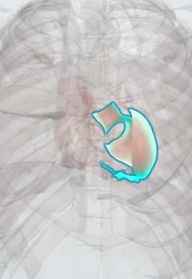
Hypoplastic left heart syndrome !
A baby is born with a tiny left heart that cannot pump to the body. It needs urgent care and several surgeries in the first years.
Updated 2026-11-01
Structures involved
Tap one to see what it is and open it in 3D.
- Left ventricle
- Mitral anterior leaflet
- Ascending aorta
- Mitral posterior leaflet
- Aortic right coronary cusp
- Aortic left coronary cusp
- Aortic non-coronary cusp
- Aortic arch
- Ligamentum arteriosum
- Interatrial septum
- Right ventricle
- Pulmonary trunk
- Superior vena cava
Why it happens
What it is
Hypoplastic left heart syndrome (HLHS) is a critical congenital heart defect in which the left-sided structures are severely underdeveloped: mitral stenosis or atresia, a small left ventricle, aortic stenosis or atresia and a hypoplastic ascending aorta and arch, often with coarctation. The right ventricle must supply both the lungs and, through the ductus arteriosus, the body, so systemic circulation is ductus-dependent.
Who gets it
About 2–3% of congenital heart disease, roughly 1 in 4,000 live births, more frequent in boys. It is one of the leading causes of death from heart disease in the first week of life. In Colombia and Latin America, outcomes depend heavily on prenatal diagnosis and timely transfer to the few centres that offer staged neonatal surgery.
Causes
- Abnormal development of the left heart in early pregnancy; the exact cause is unknown in most cases
- Reduced flow through the left heart in fetal life (for example premature restriction of the foramen ovale or fetal aortic stenosis) leads to under-growth
- Genetic associations: Turner syndrome, Jacobsen syndrome (11q deletion), trisomies 13 and 18, familial left-sided obstructive lesions
Risk factors
- Family history of left-sided obstructive heart defects (bicuspid aortic valve, coarctation)
- Chromosomal abnormalities
- Male sex
Pathophysiology
Oxygenated pulmonary venous blood cannot leave the small left heart and must cross the atrial septum to the right atrium, where it mixes with systemic venous return. The right ventricle pumps the mixed blood into the pulmonary trunk; part goes to the lungs and part reaches the aorta through the ductus arteriosus, filling the ascending aorta and coronary arteries retrogradely. When the ductus closes in the first days, systemic perfusion collapses, with shock and metabolic acidosis. A restrictive atrial septum causes severe pulmonary venous congestion and hypoxaemia at birth. Balance between pulmonary and systemic flow is delicate: falling pulmonary resistance (for example with high inspired oxygen) steals flow from the body.
Classification
- Anatomical subtypes by valve status: mitral and aortic atresia (most severe), mitral stenosis with aortic atresia, mitral and aortic stenosis
- With intact or highly restrictive atrial septum (high-risk subgroup)
Symptoms and signs
- Often well at birth, then deterioration in the first days as the ductus closes
- Tachypnoea, poor feeding and lethargy
- Grey or pale colour, cool extremities, weak or absent pulses in all limbs
- Mild cyanosis
- Shock with oliguria and metabolic acidosis
Diagnosis
- Clinical
- Many cases are diagnosed prenatally on the four-chamber view of the fetal ultrasound, confirmed by fetal echocardiography. After birth: a newborn in shock with diffusely weak pulses, single loud S2 and often no significant murmur.
- Tests
- Pulse oximetry screening; arterial blood gas and lactate (metabolic acidosis); renal and liver function; glucose. ECG shows right ventricular dominance with reduced left-sided forces.
- Imaging
- Echocardiography is diagnostic: small or slit-like left ventricle, atretic or stenotic mitral and aortic valves, small ascending aorta with retrograde flow, size of the atrial communication and ductus. Chest X-ray: cardiomegaly and pulmonary venous congestion.
Differential diagnosis
- Critical aortic stenosis or interrupted aortic arch
- Severe coarctation of the aorta
- Neonatal sepsis
- Inborn errors of metabolism presenting with shock
- Other cyanotic heart defects (transposition, TAPVR)
Goal of treatment
Keep the ductus open and balance pulmonary and systemic blood flow after birth, then convert to a single-ventricle (Fontan) circulation through staged surgery, or offer heart transplantation.
Treatment
- Acute
- Prostaglandin E1 infusion to keep the ductus arteriosus open; avoid excess supplemental oxygen and hyperventilation so that pulmonary flow does not steal from the systemic circulation; inotropes and ventilation as needed; urgent atrial septostomy if the septum is restrictive.
- Surgical
- Stage I (Norwood procedure, first week): the pulmonary trunk is used to build a new aorta, the atrial septum is opened and lung flow is supplied by a systemic-to-pulmonary shunt or a right ventricle-to-pulmonary artery conduit; a hybrid approach (ductal stent plus pulmonary artery bands) is an alternative.
- Surgical
- Stage II (bidirectional Glenn, around 3–6 months) connects the superior vena cava to the pulmonary arteries; stage III (Fontan, around 2–5 years) routes inferior vena cava blood to the lungs as well.
- Surgical
- Heart transplantation as an alternative, or when the staged circulation fails.
- Follow-up
- Nursing: apnoea and temperature monitoring on prostaglandin, oxygen saturation targets set by the team, feeding and weight gain; interstage home monitoring of saturation and weight with clear return criteria; lifelong congenital cardiology follow-up and neurodevelopmental assessment.
Complications
- Death from shock if the ductus closes before treatment
- Interstage sudden deterioration and death
- Arrhythmias and failure of the single right ventricle; tricuspid regurgitation
- Fontan complications: protein-losing enteropathy, plastic bronchitis, thrombosis, Fontan-associated liver disease
- Neurodevelopmental delay
Prognosis
Without treatment it is fatal within the first weeks of life. With staged surgery most infants survive the first stage and many now reach adulthood, but the Fontan circulation is palliative and needs lifelong specialised follow-up; some patients eventually need transplantation.
Prevention
- Prenatal ultrasound screening and fetal echocardiography to plan delivery at a cardiac surgical centre
- Universal newborn pulse oximetry screening
- Genetic counselling for families with previous left-sided heart defects
Test yourself
Think of the answer, then tap the question to see it.
On which structure does systemic blood flow depend in HLHS before surgery?
The ductus arteriosus.
Which drug is started immediately after diagnosis?
Prostaglandin E1, to keep the ductus open.
Why is high inspired oxygen avoided?
It lowers pulmonary resistance and diverts flow from the systemic circulation to the lungs.
What are the three stages of surgical palliation?
Norwood (stage I), bidirectional Glenn (stage II) and Fontan (stage III).
Which chromosomal syndrome in girls is associated with left-sided obstructive lesions including HLHS?
Turner syndrome (45,X).
Related conditions
- Coarctation of the aorta
- Turner syndrome
- Patent ductus arteriosus
- Atrial septal defect
- Heart failure
- Cardiogenic shock
- Transposition of the great arteries
- Truncus arteriosus, TAPVR and tricuspid atresia
- Neonatal sepsis
Educational information, not medical advice. For any symptom, see a health professional.
Sources: Hypoplastic left heart syndrome (MedlinePlus, US National Library of Medicine, 2026); Hypoplastic Left Heart Syndrome (HLHS) (Centers for Disease Control and Prevention (CDC), 2026).
Spotted a mistake?We are always improving for the community. Tell us what is wrong and our team will review it.Report a mistake
Representative model, for educational purposes only.