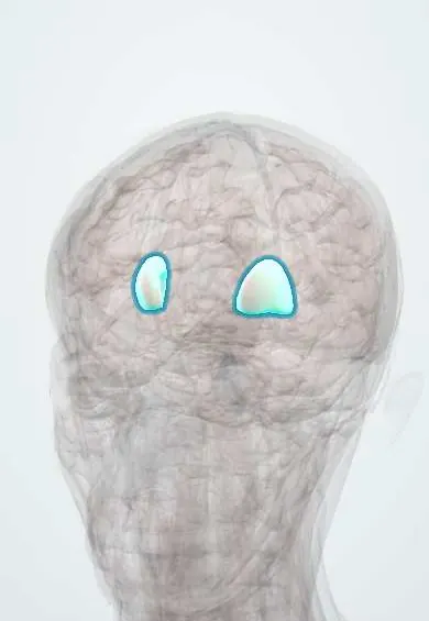
Multiple system atrophy (MSA) !
A rare, fast-progressing brain disease that combines Parkinson-like slowness or poor balance with failure of automatic functions such as blood pressure.
Updated 2026-11-01
Structures involved
Tap one to see what it is and open it in 3D.
- Putamen
- Pons
- Cerebellum
- Substantia nigra
- Caudate nucleus
- Globus pallidus
- Cerebellar peduncles
- Medulla oblongata
- Spinal cord
- Sympathetic trunk
Why it happens
- Intracellular accumulations: cholesterol, proteins, hyaline, glycogen
- Basal ganglia and movement control
- The autonomic nervous system: organization
- Glial cells: astrocytes, oligodendrocytes, microglia, Schwann cells
What it is
A sporadic, adult-onset neurodegenerative disease in which alpha-synuclein builds up inside glial cells (oligodendrocytes). It damages the basal ganglia, the cerebellum and its brainstem connections, and the autonomic centres. Patients show autonomic failure together with either parkinsonism that responds poorly to levodopa (MSA-P) or cerebellar ataxia (MSA-C). It belongs to the synucleinopathies with Parkinson disease and dementia with Lewy bodies.
Who gets it
Prevalence is roughly 2 to 5 per 100,000, with onset usually in the sixth decade (mean around 55 years). Men and women are affected about equally. The parkinsonian type predominates in Western series and the cerebellar type in Japan. There are no reliable Colombian data; in Latin America it is probably under-diagnosed and often first labelled as Parkinson disease.
Causes
- Misfolded alpha-synuclein accumulating in oligodendrocytes (glial cytoplasmic inclusions)
- Cause unknown; almost always sporadic, not inherited
- Possible genetic susceptibility and environmental factors under study
Risk factors
- Age between about 50 and 60 years
- REM sleep behaviour disorder, which often precedes motor signs by years
- No modifiable risk factor is established
Pathophysiology
Alpha-synuclein aggregates in oligodendrocytes, the cells that make central myelin, and then spreads to neurons. Neurons and myelin are lost in the striatum (especially the putamen) and substantia nigra, causing parkinsonism; in the pons, middle cerebellar peduncles and cerebellum, causing ataxia; and in the brainstem autonomic nuclei and the intermediolateral columns of the spinal cord, causing autonomic failure. Because the striatal neurons that carry dopamine receptors are themselves lost, replacing dopamine with levodopa helps little. The autonomic lesion is preganglionic, so cardiac sympathetic nerves are usually preserved, unlike in Parkinson disease.
Classification
- MSA-P: parkinsonism predominant (slowness, rigidity, falls)
- MSA-C: cerebellar ataxia predominant (gait ataxia, dysarthria, abnormal eye movements)
- Clinical certainty levels: possible, probable and clinically established; definite MSA requires neuropathology
Symptoms and signs
- Autonomic failure: orthostatic hypotension (dizziness or fainting on standing), urinary urgency, incontinence or retention, erectile dysfunction, constipation and reduced sweating
- Parkinsonism: slowness, rigidity and early falls, usually symmetric, with irregular jerky tremor rather than classic resting tremor
- Cerebellar signs: broad-based unsteady gait, limb ataxia, slurred speech and nystagmus
- Speech and swallowing problems; a soft, strained or high-pitched voice
- Breathing problems in sleep: inspiratory stridor and sleep apnoea
- Dream enactment (REM sleep behaviour disorder), cold hands, neck flexion (antecollis) and emotional incontinence
Diagnosis
- Clinical
- Suspected in an adult over 30 with progressive autonomic failure (orthostatic hypotension or urogenital dysfunction) plus parkinsonism with a poor or short-lived levodopa response, or plus cerebellar ataxia. Early severe autonomic failure, early falls, rapid progression, stridor and absence of dementia early on favour MSA over Parkinson disease.
- Tests
- Orthostatic blood pressure measurements (a fall of at least 20 mmHg systolic or 10 mmHg diastolic within 3 minutes of standing; criteria for MSA use larger falls). Bladder ultrasound for post-void residual and urodynamic studies. Autonomic function tests. Cardiac MIBG scintigraphy is usually normal (preganglionic lesion), whereas it is abnormal in Parkinson disease. Polysomnography documents REM sleep behaviour disorder and stridor. Skin biopsy or fluid alpha-synuclein assays are emerging supportive tests.
- Imaging
- Brain MRI may show atrophy of the putamen, pons, middle cerebellar peduncles and cerebellum; the "hot cross bun" sign (cross-shaped hyperintensity in the pons) in MSA-C and a hyperintense putaminal rim in MSA-P. Dopamine transporter imaging is abnormal but does not separate MSA from Parkinson disease.
Differential diagnosis
- Parkinson disease
- Dementia with Lewy bodies
- Progressive supranuclear palsy
- Spinocerebellar ataxias and other adult-onset ataxias (alcohol, vitamin deficiency)
- Pure autonomic failure
- Drug-induced orthostatic hypotension or parkinsonism
Goal of treatment
No treatment slows MSA; care aims to control orthostatic hypotension, bladder dysfunction and motor symptoms, prevent falls and aspiration, protect the airway and maintain quality of life with a multidisciplinary team.
Treatment
- Conservative
- Non-drug measures for orthostatic hypotension: liberal fluid and salt intake, compression stockings or abdominal binder, slow position changes, sleeping with the head of the bed raised and avoiding hot environments and large meals.
- Medical
- Drugs for orthostatic hypotension when non-drug measures fail: a volume expander (fludrocortisone), alpha-1 agonists (midodrine) or a norepinephrine precursor (droxidopa), watching for supine hypertension.
- Medical
- A trial of levodopa for parkinsonism (benefit is partial and short-lived in most); antimuscarinic drugs for overactive bladder; treatment of constipation and erectile dysfunction.
- Procedure
- Intermittent clean catheterisation for urinary retention; continuous positive airway pressure or, for severe stridor, tracheostomy; gastrostomy when swallowing is unsafe.
- Rehabilitation
- Physiotherapy for gait and fall prevention, speech and swallowing therapy, occupational therapy and mobility aids.
- Follow-up
- Nursing priorities: lying and standing blood pressure, fall-risk assessment, swallowing screening, post-void residual, skin integrity and palliative-care planning.
Complications
- Falls and fractures
- Aspiration pneumonia
- Sudden death in sleep from stridor or central apnoea
- Urinary tract infections from retention
- Pressure ulcers and immobility
- Supine hypertension caused by treatment
Prognosis
Progression is faster than in Parkinson disease: many patients need a wheelchair within about 5 years, and median survival is roughly 6 to 10 years from symptom onset. Death usually results from aspiration pneumonia, other infections or sudden death in sleep.
Test yourself
Think of the answer, then tap the question to see it.
What is the hallmark inclusion of multiple system atrophy?
Alpha-synuclein glial cytoplasmic inclusions in oligodendrocytes.
Which combination of features suggests MSA rather than Parkinson disease?
Early severe autonomic failure (orthostatic hypotension, urinary dysfunction) with parkinsonism that responds poorly to levodopa or with cerebellar ataxia.
What MRI sign is classic for the cerebellar type of MSA?
The "hot cross bun" sign in the pons, with atrophy of the pons and middle cerebellar peduncles.
Why does cardiac MIBG scintigraphy help separate MSA from Parkinson disease?
In MSA the autonomic lesion is preganglionic, so cardiac sympathetic uptake is usually normal; in Parkinson disease it is reduced.
Which sleep-related sign in MSA is dangerous and needs urgent attention?
Inspiratory stridor, which can cause sudden death in sleep.
Related conditions
- Parkinson disease
- Dementia with Lewy bodies
- Progressive supranuclear palsy
- REM sleep behaviour disorder (RBD)
- Spinocerebellar ataxias (autosomal dominant ataxias)
- Syncope (vasovagal, orthostatic hypotension, cardiac)
- Neurogenic bladder
- Erectile dysfunction
- Obstructive sleep apnoea
Educational information, not medical advice. For any symptom, see a health professional.
Sources: Multiple system atrophy: MedlinePlus Genetics (MedlinePlus (U.S. National Library of Medicine), 2026); Multiple System Atrophy - Neurology - MSD Manual Professional Edition (MSD Manual Professional Edition, 2026).
Spotted a mistake?We are always improving for the community. Tell us what is wrong and our team will review it.Report a mistake
Representative model, for educational purposes only.