Conditions · Emergency medicine and toxicology
Neurogenic shock !
Low blood pressure with a slow pulse after a high spinal cord injury, because the nerves that keep vessels tight and the heart fast stop working.
Updated 2026-11-01
Structures involved
Tap one to see what it is and open it in 3D.
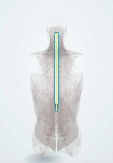
- Spinal cord
- Inferior vena cava
- Sympathetic trunk
- Vagus nerve
- Grey matter of the cord
- Neck nerve roots
- Mid-back nerve roots
- C5 vertebra
- T6 vertebra
- Thoracic aorta
Why it happens
- Shock: types, stages and the organs it damages
- Shock physiology
- The autonomic nervous system: organization
- Nervous control of the circulation and the CNS ischemic response
What it is
A form of distributive shock caused by loss of sympathetic tone after an injury to the cervical or upper thoracic spinal cord (usually above T6). Vessels dilate and the heart is driven only by the vagus nerve, giving hypotension with bradycardia and warm, dry skin.
Who gets it
Seen mainly after cervical spinal cord injury from road traffic collisions, especially motorcycle crashes, falls, violence and diving in shallow water, all common causes of trauma in Colombia. It appears in a sizeable share of cervical cord injuries and is less frequent in lower injuries.
Causes
- Traumatic cervical or high thoracic spinal cord injury
- Less often: high spinal or epidural anaesthesia, transverse myelitis or cord compression
Risk factors
- Complete cord injury
- Injury level at or above T6, especially cervical
- High-energy trauma: motorcycle crashes, falls from height, diving into shallow water, gunshot wounds
Pathophysiology
Sympathetic outflow leaves the cord from T1 to L2. A high cord injury cuts the descending pathways to it, so arterial and venous tone fall (low resistance and pooled venous return) and the heart loses sympathetic drive (T1 to T4) while vagal input is preserved. The result is low blood pressure with bradycardia, poor cardiac output and loss of temperature control.
Classification
- Neurogenic shock: haemodynamic (hypotension, bradycardia, vasodilation)
- Spinal shock: temporary loss of all cord function, reflexes and tone below the injury; neurological, not haemodynamic
Symptoms and signs
- Hypotension
- Bradycardia or a heart rate that fails to rise despite low pressure
- Warm, dry, pink skin below the injury (unlike the cold clammy skin of hypovolaemic shock)
- Flaccid paralysis and loss of sensation below the level of injury
- Priapism, loss of rectal tone and poor temperature control (hypothermia)
- Breathing difficulty in high cervical injuries
Diagnosis
- Clinical
- Diagnosis of exclusion in a trauma patient with a cord injury: hypotension with bradycardia and warm skin, after ruling out bleeding, tension pneumothorax and tamponade. Full neurological examination (ASIA/ISNCSCI scale) to set the level.
- Tests
- Continuous ECG and blood pressure monitoring, arterial blood gas and lactate, haemoglobin; FAST ultrasound to look for internal bleeding.
- Imaging
- CT of the spine to find fractures; MRI to show cord and ligament damage; chest X-ray and abdominal imaging to rule out other causes of shock.
Differential diagnosis
- Hypovolaemic (haemorrhagic) shock, the most frequent cause of shock in trauma
- Obstructive shock: tension pneumothorax or cardiac tamponade
- Cardiogenic shock from blunt cardiac injury
- Septic shock or anaphylaxis
- Spinal shock without haemodynamic change
Goal of treatment
Restore perfusion of the injured cord and organs, avoid hypotension and hypoxia, and protect the spine from further injury.
Treatment
- Acute
- ATLS primary survey with airway protection (early intubation in high cervical injuries) and spinal motion restriction; bleeding is sought and controlled first.
- Medical
- Careful intravenous crystalloids, then vasopressors with alpha and beta effect (norepinephrine) or alpha agonists (phenylephrine, less suitable if bradycardic) to keep a target mean arterial pressure, often 85 to 90 mmHg in the first days.
- Medical
- Anticholinergics (atropine) for symptomatic bradycardia; temporary pacing if it persists.
- Surgical
- Spinal decompression and stabilisation by the spine team when indicated.
- Follow-up
- ICU care: invasive blood pressure, heart rhythm, urine output and temperature; nursing care for pressure areas, bladder, bowel and venous thromboembolism prophylaxis.
Complications
- Secondary cord ischaemia and worse neurological outcome from hypotension
- Bradyarrhythmias and cardiac arrest
- Respiratory failure and pneumonia
- Pressure injuries, deep vein thrombosis and pulmonary embolism
- Later autonomic dysreflexia in injuries above T6
Prognosis
The haemodynamic instability usually improves over days to weeks as reflex vascular tone returns. The final outcome depends on the level and completeness of the cord injury; avoiding hypotension early improves neurological recovery.
Test yourself
Think of the answer, then tap the question to see it.
What combination of vital signs suggests neurogenic shock?
Hypotension with bradycardia (or a normal rate) and warm, dry skin after a high spinal cord injury.
Above which spinal level does neurogenic shock usually occur?
Above T6, mostly in cervical injuries, because sympathetic outflow (T1 to L2) and cardiac accelerator fibres (T1 to T4) are cut off.
How does neurogenic shock differ from spinal shock?
Neurogenic shock is haemodynamic (hypotension, bradycardia); spinal shock is a temporary loss of reflexes, tone and sensation below the injury.
What must be ruled out first in a hypotensive patient with a spinal cord injury?
Haemorrhagic (hypovolaemic) shock, the most common cause of shock in trauma, which can coexist.
Which drug classes are used when fluids do not correct neurogenic shock?
Vasopressors such as norepinephrine or phenylephrine, and anticholinergics (atropine) for symptomatic bradycardia.
Related conditions
- Spinal cord injury
- Cervical spine fracture
- Hypovolaemic shock
- Sepsis and septic shock
- Cardiogenic shock
- Anaphylaxis
- Autonomic dysreflexia
- Traumatic brain injury
- Deep vein thrombosis
- Pressure injury (pressure ulcer)
Educational information, not medical advice. For any symptom, see a health professional.
Sources: Shock (MedlinePlus, US National Library of Medicine, 2026); Spinal cord trauma (MedlinePlus, US National Library of Medicine, 2026).
Spotted a mistake?We are always improving for the community. Tell us what is wrong and our team will review it.Report a mistake
Representative model, for educational purposes only.