Conditions · Surgery and trauma
Cervical spine fracture !
A cervical spine fracture is a break in one of the seven neck bones. It can injure the spinal cord, so the neck is kept still until it is cleared.
Updated 2026-11-01
Structures involved
Tap one to see what it is and open it in 3D.
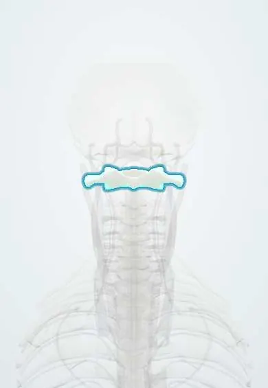
- Atlas (C1)
- Axis (C2)
- C7 vertebra
- Spinal cord
- C3 vertebra
- C4 vertebra
- C5 vertebra
- C6 vertebra
- Vertebral artery
- Anterior longitudinal ligament
- Posterior longitudinal ligament
- Ligamentum flavum
- Interspinous ligaments
- Neck facet joints
- C5–C6 disc
- Neck nerve roots
Why it happens
- Upper versus lower motor neuron lesions
- Somatosensory pathways: dorsal column and anterolateral
- The autonomic nervous system: organization
What it is
A fracture or ligament-unstable injury of one or more of the seven cervical vertebrae (C1–C7). Its importance lies in the risk to the spinal cord and the vertebral arteries; until injury is excluded, every major trauma patient is treated as if the neck were broken.
Who gets it
Most injuries follow high-energy blunt trauma in young adults, and low falls in older adults. In Colombia and Latin America, motorcycle crashes, falls from height and diving into shallow water are typical mechanisms. Clinically important neck injury is found in only a small fraction of blunt trauma patients assessed, which is why decision rules are used to limit imaging.
Causes
- Road traffic crashes, especially motorcycle riders and occupants without seatbelts
- Falls from height, and ground-level falls in older adults
- Diving into shallow water (axial loading)
- Contact sports and horse riding
- Assaults and gunshot wounds
Risk factors
- High-energy mechanism or associated head injury
- Age over 65 years
- Osteoporosis
- Ankylosing spondylitis or diffuse spinal ossification (rigid spine that breaks like a long bone)
- Rheumatoid arthritis or Down syndrome (atlantoaxial instability)
- Alcohol intoxication
Pathophysiology
Flexion, extension, axial compression or rotation forces break the bone, tear the stabilising ligaments, or both. Instability lets the fragments or the displaced vertebrae compress or tear the spinal cord, nerve roots or vertebral arteries. Injury above C5 can paralyse the diaphragm, and loss of sympathetic tone above T6 can cause neurogenic shock with hypotension and bradycardia.
Classification
- Jefferson fracture: burst fracture of the C1 ring from axial loading
- Odontoid (dens) fractures of C2: types I, II and III (type II is the most unstable and the most prone to nonunion)
- Hangman's fracture: bilateral pars fracture of C2 from hyperextension
- Subaxial C3–C7 injuries: compression, burst, flexion teardrop, facet dislocation and clay-shoveler's (C7 spinous process) fractures
- Stable versus unstable, and with or without neurological deficit
Symptoms and signs
- Neck pain and midline tenderness over the spinous processes
- Muscle spasm and reluctance to move the head
- Numbness, tingling or weakness in the arms or legs
- Pain radiating into the shoulders or arms
- Breathing difficulty in high cervical injuries
Diagnosis
- Clinical
- Spinal motion restriction from the scene, then ATLS primary survey and a full neurological examination with sensory level. Validated rules decide who needs imaging: NEXUS (no midline tenderness, no focal deficit, normal alertness, no intoxication, no painful distracting injury) and the Canadian C-spine rule (high-risk factors, low-risk factors allowing assessment, and the ability to rotate the neck 45° each way).
- Tests
- Baseline blood tests as part of trauma care; arterial blood gas and vital capacity monitoring in high cervical injuries.
- Imaging
- CT of the cervical spine is the first study in adults who need imaging and has largely replaced plain films. MRI shows the cord, discs and ligaments when there is a deficit or suspected ligament injury; CT angiography assesses the vertebral arteries in high-risk fracture patterns.
Differential diagnosis
- Cervical muscle strain or whiplash injury
- Isolated ligament injury without fracture
- Acute disc herniation
- Spinal cord injury without radiographic abnormality, especially in children
- Pre-existing degenerative changes mistaken for acute injury
Goal of treatment
Protect the spinal cord from further harm, restore alignment and stability, and support breathing and circulation while the injury heals.
Treatment
- Acute
- ATLS approach with spinal motion restriction, in-line stabilisation during airway management, oxygen, and treatment of hypotension; log-roll for examination and transfers.
- Conservative
- Stable fractures without deficit are treated with a rigid cervical collar or brace for several weeks, with follow-up imaging.
- Procedure
- Closed reduction with skull traction for some dislocations, and halo-vest immobilisation for selected upper cervical fractures.
- Surgical
- Decompression and fusion with internal fixation for unstable fractures, dislocations and injuries with cord compression or progressive deficit.
- Follow-up
- Nursing priorities: airway and respiratory function, repeated neurological checks, blood pressure and heart rate (neurogenic shock), pressure-injury prevention, bladder care, and pin-site and collar skin care.
- Rehabilitation
- Physiotherapy and, after cord injury, specialised spinal rehabilitation.
Complications
- Spinal cord injury with quadriplegia
- Respiratory failure and pneumonia
- Neurogenic shock
- Vertebral artery injury and posterior circulation stroke
- Nonunion (especially type II odontoid fractures) and late instability
- Pressure injuries and venous thromboembolism from immobility
Prognosis
Stable fractures without neurological deficit usually heal well with bracing. Outcome after cord involvement depends on the level and completeness of the cord injury; older adults with upper cervical fractures have higher mortality.
Prevention
- Seatbelts, child restraints and motorcycle safety
- Avoiding diving into shallow or unknown water
- Fall prevention in older adults
- Correct technique and protective equipment in contact sports
Test yourself
Think of the answer, then tap the question to see it.
Which C2 fracture results from hyperextension and involves both pars interarticularis?
The hangman's fracture (traumatic spondylolisthesis of the axis).
What is the first-line imaging study for an adult with a suspected cervical spine fracture after blunt trauma?
CT of the cervical spine; MRI is added for neurological deficit or suspected ligament injury.
What are the five NEXUS criteria that allow clinical clearance of the cervical spine?
No midline tenderness, no focal neurological deficit, normal alertness, no intoxication and no painful distracting injury.
Hypotension with bradycardia after a cervical spine injury suggests what?
Neurogenic shock from loss of sympathetic tone.
Which axial-loading fracture bursts the ring of C1?
The Jefferson fracture.
Related conditions
- Spinal cord injury
- Major trauma (polytrauma): ATLS primary survey
- Traumatic brain injury
- Whiplash (cervical sprain)
- Neurogenic shock
- Ankylosing spondylitis
- Rheumatoid arthritis
- Osteoporotic vertebral compression fracture
Educational information, not medical advice. For any symptom, see a health professional.
Sources: Cervical Fracture (Broken Neck) - OrthoInfo - AAOS (American Academy of Orthopaedic Surgeons (OrthoInfo), 2026); Advanced Trauma Life Support | ACS (American College of Surgeons, 2025).
Spotted a mistake?We are always improving for the community. Tell us what is wrong and our team will review it.Report a mistake
Representative model, for educational purposes only.