
Pulmonary alveolar proteinosis
A rare disease in which surfactant piles up in the air sacs because the cells that clear it fail. It causes slow breathlessness; washing out the lungs helps.
What it is
A rare syndrome in which surfactant lipids and proteins accumulate in the alveoli because alveolar macrophages cannot clear them, filling the air spaces and impairing gas exchange. Most cases are autoimmune, caused by antibodies against granulocyte-macrophage colony-stimulating factor (GM-CSF).
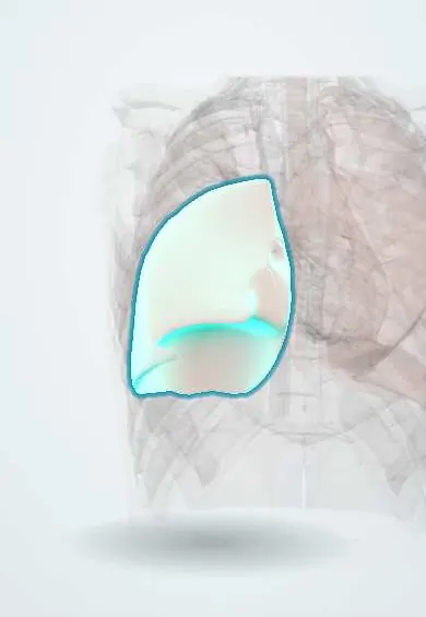
Who gets it
Rare, with a prevalence of a few cases per million people. About 90% are autoimmune; it usually appears between 30 and 50 years of age, is more frequent in men, and most patients have smoked. Secondary forms occur with blood cancers or heavy dust exposure, and hereditary forms mainly in infants and children.
Causes
- Autoimmune: neutralising anti-GM-CSF autoantibodies (most cases)
- Secondary: blood disorders (myelodysplastic syndrome, myeloid leukaemia), GATA2 deficiency, immunodeficiency and immunosuppression
- Secondary: heavy inhalation of silica (acute silicoproteinosis), aluminium, indium, titanium or other dusts and fumes
- Hereditary: mutations in the GM-CSF receptor (CSF2RA, CSF2RB)
- Congenital surfactant production disorders (SFTPB, SFTPC, ABCA3, NKX2-1), which are a different, mainly paediatric disease that looks similar
Risk factors
- Smoking
- Occupational dust and fume exposure (sandblasting, mining, stone cutting, metalworking)
- Myelodysplastic syndrome and other myeloid disorders
- Male sex, middle age
Pathophysiology
Type II pneumocytes secrete surfactant continuously → alveolar macrophages normally take up and break down most of it, but only while GM-CSF, acting through the transcription factor PU.1, keeps them mature → in autoimmune disease antibodies neutralise GM-CSF; in hereditary disease its receptor is defective; in secondary disease the macrophages are damaged or too few → surfactant piles up, macrophages become swollen and foamy, and alveoli fill with periodic acid-Schiff (PAS)-positive material while the interstitium stays largely normal → blood passing the filled alveoli is not oxygenated (shunt), so hypoxaemia and a low diffusing capacity follow → the same failing macrophages and neutrophils fight microbes poorly, so Nocardia, mycobacteria and fungi take hold.
Classification
- Primary: autoimmune (anti-GM-CSF antibodies) and hereditary (GM-CSF receptor defects)
- Secondary: haematological, inhaled dust, infection, immunodeficiency
- Congenital surfactant production disorders (separate category, mostly children)
- By severity: from radiographic disease without symptoms to hypoxaemic respiratory failure
Symptoms and signs
- Slowly progressive breathlessness on exertion
- Dry cough, sometimes with whitish or gummy sputum
- Fatigue and weight loss
- Some people have no symptoms despite marked radiographic changes
- Fever suggests a superimposed infection
- Few signs: crackles are uncommon; cyanosis and clubbing in advanced disease
Diagnosis
- Clinical
- Suspected in a middle-aged person, often a smoker, with slowly progressive breathlessness and diffuse opacities that look worse on imaging than the symptoms suggest; occupational exposures and blood disorders are asked about.
- Tests
- Serum anti-GM-CSF antibody testing is highly sensitive and specific for autoimmune proteinosis; when negative, serum GM-CSF levels and genetic testing look for hereditary forms. Bronchoalveolar lavage returns milky, opaque fluid with PAS-positive material and foamy macrophages; transbronchial or cryobiopsy, or rarely surgical biopsy, is needed only when the diagnosis is unclear. Pulmonary function shows a disproportionate fall in diffusing capacity and often a restrictive pattern; oximetry or arterial blood gas measures hypoxaemia. Serum LDH and other markers (KL-6, CEA) are often raised; a blood count screens for a haematological cause; cultures look for infection.
- Imaging
- Chest radiograph shows bilateral, symmetrical, mainly perihilar airspace opacities (bat-wing pattern) without effusions or cardiomegaly. High-resolution CT shows ground-glass opacity with thickened interlobular septa (crazy paving) and sharp, geographic borders with spared lobules.
Differential diagnosis
- Cardiogenic pulmonary oedema
- Pneumocystis pneumonia and other atypical infections
- Diffuse alveolar haemorrhage
- Acute respiratory distress syndrome
- Organizing pneumonia and non-specific interstitial pneumonia
- Lipoid pneumonia
- Invasive mucinous adenocarcinoma
- Sarcoidosis
Bilateral ground glass and crazy paving: alveolar proteinosis and its mimics
| Condition | Course | Distinguishing finding | Bronchoalveolar lavage |
|---|---|---|---|
| Pulmonary alveolar proteinosis | Slow, over months; symptoms milder than the imaging | Geographic crazy paving with spared lobules; anti-GM-CSF antibodies | Milky, opaque; PAS-positive material and foamy macrophages |
| Pneumocystis pneumonia | Subacute, days to weeks; fever, oxygen falls on exertion | Immunosuppression (HIV, corticosteroids), high LDH | Organisms on silver stain or PCR |
| Cardiogenic pulmonary oedema | Hours to days | Large heart, effusions, septal lines, quick response to diuretics | Rarely needed |
| Diffuse alveolar haemorrhage | Acute; haemoptysis and falling haemoglobin | Vasculitis or kidney involvement | Increasingly bloody returns; haemosiderin-laden macrophages |
Goal of treatment
Clear the alveoli to restore gas exchange, correct the underlying cause of macrophage failure, prevent and treat infection, and avoid further exposure.
Treatment
- Follow-up
- People with mild disease and few symptoms are followed with symptoms, oximetry, lung function and imaging; some remit spontaneously.
- Procedure
- Whole-lung lavage, the standard treatment for symptomatic disease: under general anaesthesia with a double-lumen tube, one lung is ventilated while the other is repeatedly filled with warmed saline and drained, with chest percussion, until the fluid clears; the other lung follows in the same or a later session.
- Medical
- Inhaled recombinant GM-CSF (sargramostim, molgramostim) for autoimmune proteinosis improves oxygenation and may reduce the need for lavage; availability varies by country. Rituximab or plasmapheresis has been tried in refractory cases with limited evidence.
- Medical
- Secondary forms: treat the cause, for example removal from dust exposure or treatment of the blood disorder; haematopoietic stem cell transplantation for GATA2 deficiency or hereditary receptor defects in selected patients.
- Lifestyle
- Stop smoking and avoid occupational dusts; vaccination against influenza and pneumococcus; prompt treatment of infections.
- Surgical
- Lung transplantation for end-stage disease or congenital surfactant disorders; the autoimmune form can recur in the transplanted lung.
Complications
- Hypoxaemic respiratory failure
- Opportunistic infections: Nocardia, Mycobacterium (tuberculous and non-tuberculous), Aspergillus, Cryptococcus, Histoplasma
- Pulmonary fibrosis (uncommon)
- Complications of lavage: transient worse hypoxaemia, fluid leak into the pleura, pneumothorax
Common traps
- Alveolar proteinosis is a disease of surfactant overproduction: in adults production is normal; clearance fails because the macrophages lack GM-CSF signalling
- Crazy paving on CT means alveolar proteinosis: it also appears with pulmonary oedema, Pneumocystis, alveolar haemorrhage, ARDS and mucinous adenocarcinoma; anti-GM-CSF antibodies and lavage confirm it
- Fever is part of the disease: proteinosis itself rarely causes fever; fever points to an opportunistic infection such as Nocardia, mycobacteria or fungi
- Imaging this extensive means the patient must be very ill: symptoms are often milder than the CT suggests, and some patients have none
- Congenital surfactant gene defects are the childhood form of the same disease: SFTPB, SFTPC, ABCA3 and NKX2-1 defects disturb surfactant production, a different disease that only looks alike
Prognosis
Autoimmune proteinosis has a good outlook with lavage and GM-CSF, with five-year survival above 90%; some cases remit on their own and others need repeated lavage. Secondary proteinosis depends on its cause and is worse with blood cancers; congenital surfactant disorders are often severe in infants.
Test yourself
Think of the answer, then tap the question to see it.
What causes most cases of pulmonary alveolar proteinosis?
Autoantibodies that neutralise GM-CSF, so alveolar macrophages cannot mature and clear surfactant.
What is the classic high-resolution CT pattern?
Crazy paving: ground-glass opacity with thickened interlobular septa, with sharp geographic borders and spared lobules.
What does bronchoalveolar lavage look like in this disease?
Milky, opaque fluid with PAS-positive acellular material and foamy macrophages.
What is the standard treatment for symptomatic disease?
Whole-lung lavage under general anaesthesia; inhaled GM-CSF is an option in the autoimmune form.
A 42-year-old smoker has 8 months of worsening exertional breathlessness, no fever. CT: bilateral ground glass, thickened septa, sharp geographic borders. Which blood test is most useful next?
Serum anti-GM-CSF antibodies: highly sensitive and specific for autoimmune alveolar proteinosis, the likely diagnosis given the crazy paving and the slow course without fever.
Why it happens
Connections
Educational information, not medical advice. For any symptom, see a health professional.
Sources: Pulmonary Alveolar Proteinosis (StatPearls (NCBI Bookshelf), 2026); Bronchoalveolar Lavage (StatPearls (NCBI Bookshelf), 2024).
Spotted a mistake?We are always improving for the community. Tell us what is wrong and our team will review it.Report a mistake
Representative model, for educational purposes only.