
The lung transplant recipient
Life after receiving a new lung or lungs: lifelong anti-rejection medicines and close follow-up to catch rejection, infection and drug side effects early.
What it is
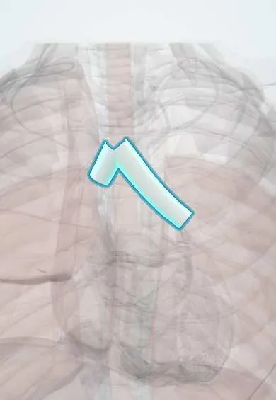
A person living with one or two transplanted lungs for end-stage lung disease. Care centres on lifelong immunosuppression and close monitoring, because the lung has the highest rates of rejection and infection among transplanted solid organs.
Who gets it
About 4,500 to 5,000 lung transplants are done worldwide each year, most of them bilateral. The main indications are interstitial lung disease (especially idiopathic pulmonary fibrosis), chronic obstructive pulmonary disease, cystic fibrosis and pulmonary arterial hypertension. Median survival is about 6 to 7 years, longer after bilateral than single transplants.
Causes
- Why it is done: interstitial lung disease, especially idiopathic pulmonary fibrosis
- Chronic obstructive pulmonary disease, including alpha-1 antitrypsin deficiency
- Cystic fibrosis and other bronchiectasis
- Pulmonary arterial hypertension (sometimes heart-lung transplant)
- Less often: sarcoidosis, lymphangioleiomyomatosis, retransplantation for chronic graft dysfunction
Risk factors
- Primary graft dysfunction in the first 72 hours
- Older recipient age and frailty
- Cytomegalovirus mismatch (seropositive donor, seronegative recipient)
- Gastro-oesophageal reflux and silent aspiration
- Donor-specific antibodies and repeated acute rejection
- Respiratory viral infections
- Missed doses or poor adherence to immunosuppression
Pathophysiology
The donor lung is joined at three sites (main bronchus, pulmonary artery and a left atrial cuff carrying the pulmonary veins), but its bronchial arteries, vagal nerves and lymphatics are cut → the donor bronchus depends on collateral flow and may become ischaemic (stenosis, dehiscence), and without vagal fibres there is no cough below the anastomosis, so secretions and aspirated reflux stay in the graft → constant exposure to inhaled microbes and particles keeps the immune system activated → recipient T cells attack donor tissue around vessels and small airways (acute cellular rejection), and donor-specific antibodies injure capillaries (antibody-mediated rejection) → repeated injury from rejection, infection and reflux scars the graft: fibrous closure of small airways (bronchiolitis obliterans syndrome) or pleural and parenchymal fibrosis (restrictive allograft syndrome), together called chronic lung allograft dysfunction.
Classification
- By procedure: single, bilateral (sequential) or heart-lung transplant
- By time: first month (surgical problems, primary graft dysfunction, bacterial infection), 1-6 months (opportunistic infection such as cytomegalovirus and Pneumocystis, acute rejection), after 6 months (chronic dysfunction, community infections, cancer, kidney disease)
- Rejection types: hyperacute (rare), acute cellular (graded A0-A4 and B by biopsy), antibody-mediated, and chronic lung allograft dysfunction
- Chronic lung allograft dysfunction phenotypes: bronchiolitis obliterans syndrome (obstructive), restrictive allograft syndrome, mixed
Symptoms and signs
- Often silent: a fall in home spirometry may be the first sign
- Breathlessness, cough and reduced exercise tolerance
- Fever, sputum or flu-like illness (infection or acute rejection)
- Drug effects: tremor, high blood pressure, high blood sugar, kidney impairment, diarrhoea
- Lumps, swollen nodes or skin lesions (lymphoproliferative disorder, skin cancer)
Diagnosis
- Clinical
- Any new symptom is assessed by the transplant team, considering the time since transplant, recent changes in immunosuppression, adherence and exposure to infection. Rejection and infection often look alike and may coexist.
- Tests
- Spirometry at home and in clinic compared with the best post-transplant FEV1. Bronchoscopy with bronchoalveolar lavage and transbronchial biopsies (surveillance or for cause) grades acute rejection, finds infection and shows the anastomosis. Donor-specific antibodies; cytomegalovirus and Epstein-Barr virus PCR; calcineurin inhibitor trough levels; kidney function, glucose, lipids and blood counts. Donor-derived cell-free DNA in blood is an emerging marker of graft injury.
- Imaging
- Chest radiograph and high-resolution CT with expiratory images: air trapping and bronchial wall thickening in bronchiolitis obliterans syndrome, upper-lobe pleural and parenchymal fibrosis in restrictive allograft syndrome, consolidation or nodules in infection or lymphoproliferative disorder. Bone densitometry and age-appropriate cancer screening during follow-up.
Differential diagnosis
- Acute cellular rejection
- Antibody-mediated rejection
- Infection: bacterial, respiratory viruses, cytomegalovirus, Aspergillus, Pneumocystis, mycobacteria
- Bronchial anastomotic stenosis or dehiscence
- Aspiration and reflux
- Chronic lung allograft dysfunction
- Drug-induced pneumonitis (for example mTOR inhibitors)
- Recurrence of the original disease (sarcoidosis, lymphangioleiomyomatosis), post-transplant lymphoproliferative disorder, pulmonary embolism or heart failure
New breathlessness after lung transplant: what is it?
| Problem | Usual timing | Clues | How it is confirmed |
|---|---|---|---|
| Primary graft dysfunction | First 72 hours | Hypoxaemia and bilateral infiltrates with no other cause | Oxygenation and chest imaging criteria |
| Acute cellular rejection | Mostly the first year | Fall in FEV1, low-grade fever, infiltrates | Transbronchial biopsy (graded A and B) |
| Opportunistic infection (cytomegalovirus, fungi, Pneumocystis) | Months 1-6, or after prophylaxis stops | Fever, viraemia, nodules or ground glass | Lavage cultures and PCR, blood cytomegalovirus PCR |
| Chronic lung allograft dysfunction | After the first year | Gradual, sustained FEV1 fall; air trapping (bronchiolitis obliterans) or upper-lobe fibrosis (restrictive form) | Spirometry and CT once other causes are excluded |
Goal of treatment
Keep the graft working and the person well by balancing immunosuppression (enough to prevent rejection, little enough to limit infection and toxicity), detecting problems early and preventing infection and long-term complications.
Treatment
- Medical
- Lifelong maintenance immunosuppression, usually triple therapy: a calcineurin inhibitor (tacrolimus or ciclosporin) with drug-level monitoring, an antiproliferative agent (mycophenolate or azathioprine) and a corticosteroid; many centres add induction with basiliximab or antithymocyte globulin at transplant.
- Medical
- Infection prophylaxis: trimethoprim-sulfamethoxazole against Pneumocystis, valganciclovir or monitoring for cytomegalovirus according to donor and recipient serostatus, and antifungal prophylaxis per centre protocol; inactivated vaccines (influenza, pneumococcus, COVID-19), no live vaccines.
- Acute
- Acute cellular rejection: high-dose intravenous corticosteroid pulses; antibody-mediated rejection: plasmapheresis, intravenous immunoglobulin and, in some centres, rituximab or other agents.
- Medical
- Chronic lung allograft dysfunction: azithromycin trial, adjustment of immunosuppression, extracorporeal photopheresis in selected patients; treatment of reflux.
- Procedure
- Bronchoscopic balloon dilation, laser or stenting for bronchial anastomotic stenosis.
- Surgical
- Antireflux surgery (fundoplication) for reflux with aspiration; retransplantation for selected people with chronic graft dysfunction.
- Rehabilitation
- Pulmonary rehabilitation before and after transplantation to rebuild strength and exercise capacity.
- Follow-up
- Lifelong transplant clinic follow-up: home spirometry, drug levels, kidney function, blood pressure, glucose and lipids, bone health, and yearly skin examination.
- Lifestyle
- Strict adherence and timing of medicines, no smoking, sun protection, food and water safety, hand hygiene, avoiding sick contacts and checking any new medicine or herbal product with the team (grapefruit raises tacrolimus levels).
Complications
- Primary graft dysfunction and surgical complications (bleeding, anastomotic dehiscence or stenosis, phrenic nerve injury)
- Infections: bacterial, viral (cytomegalovirus, respiratory viruses), fungal (Aspergillus) and Pneumocystis
- Acute rejection and chronic lung allograft dysfunction
- Chronic kidney disease from calcineurin inhibitors
- Diabetes, hypertension, high cholesterol and osteoporosis
- Cancer: skin squamous cell carcinoma (worse with long-term voriconazole) and Epstein-Barr virus-related lymphoproliferative disorder
- Neurotoxicity: tremor, headache, posterior reversible encephalopathy syndrome
Common traps
- No symptoms and a clear chest X-ray mean the graft is fine: rejection and early chronic dysfunction are often silent; a sustained fall of 10% or more in FEV1 on home spirometry is the alarm
- Fever with a new infiltrate after transplant is infection: rejection and infection look alike and can coexist; bronchoscopy with lavage and biopsy separates them before immunosuppression is changed
- Any antibiotic or antifungal can be added safely: azoles and macrolides inhibit CYP3A4 and raise tacrolimus levels, and rifampicin lowers them; check interactions and drug levels with the team
- The transplanted lung coughs normally: it is denervated below the anastomosis, so cough clearance is poor and silent aspiration is common
- Chronic rejection is a single disease: chronic lung allograft dysfunction has an obstructive phenotype (bronchiolitis obliterans syndrome) and a restrictive one with a worse outlook
Prognosis
Median survival is about 6 to 7 years, better after bilateral transplant and in younger recipients; survival at one year is about 85%. Infection and primary graft dysfunction cause most early deaths, and chronic lung allograft dysfunction, present in about half of recipients by five years, causes most late deaths.
Test yourself
Think of the answer, then tap the question to see it.
Which three anastomoses are made in a lung transplant?
The main bronchus, the pulmonary artery, and a cuff of left atrium carrying the pulmonary veins.
Why is the bronchial anastomosis prone to ischaemia?
The bronchial arteries are usually not reconnected, so the donor bronchus relies on retrograde collateral flow from the pulmonary circulation.
What are the two main phenotypes of chronic lung allograft dysfunction?
Bronchiolitis obliterans syndrome (obstructive, small-airway fibrosis) and restrictive allograft syndrome (pleural and parenchymal fibrosis).
Why must azole antifungals be used with care with tacrolimus?
They inhibit CYP3A4, raising tacrolimus levels and the risk of kidney injury and neurotoxicity; levels and doses must be adjusted.
Fourteen months after a lung transplant, a woman's home FEV1 has fallen 15% over 6 weeks, without fever; CT shows expiratory air trapping. What is likely, and what must be excluded first?
Chronic lung allograft dysfunction, bronchiolitis obliterans phenotype. First exclude treatable causes (infection, acute rejection, anastomotic stenosis, reflux with aspiration), usually by bronchoscopy with lavage and biopsy.
Why it happens
In emergency medicine
Connections
Educational information, not medical advice. For any symptom, see a health professional.
Sources: Lung Transplantation (StatPearls (NCBI Bookshelf), 2024); Lung Transplant Rejection (StatPearls (NCBI Bookshelf), 2022).
Spotted a mistake?We are always improving for the community. Tell us what is wrong and our team will review it.Report a mistake
Representative model, for educational purposes only.