
Smoking-related interstitial lung disease (RB-ILD, DIP, pulmonary Langerhans cell histiocytosis)
A group of lung diseases caused by cigarette smoke that inflames the small airways and air sacs and can form cysts. Quitting smoking is the key treatment.
What it is
A group of diffuse lung diseases strongly linked to cigarette smoking: respiratory bronchiolitis–associated interstitial lung disease (RB-ILD), desquamative interstitial pneumonia (DIP) and pulmonary Langerhans cell histiocytosis (PLCH). Smoking also adds to the risk of idiopathic pulmonary fibrosis and combined pulmonary fibrosis and emphysema.
Who gets it
RB-ILD and DIP mainly affect current smokers aged 30 to 50; DIP is more common in men. Adult PLCH occurs almost only in young smokers, typically aged 20 to 40, equally in men and women. All are rare, and their true frequency is probably underestimated because they are confused with emphysema or other cystic diseases.
Causes
- Cigarette smoking in almost all cases of RB-ILD and PLCH and in most cases of DIP
- Cannabis and other inhaled smoke in some patients
- DIP without smoking: occupational dusts, drugs, infections, connective tissue diseases and inherited surfactant disorders
- PLCH: clonal proliferation of Langerhans cells, often with BRAF V600E or other MAPK-pathway mutations, triggered by smoke
Risk factors
- Current smoking, especially heavy smoking
- Young to middle adult age
- Second-hand or cannabis smoke
- Occupational dust exposure
Pathophysiology
Inhaled smoke → pigmented (smoker's) macrophages gather in the respiratory bronchioles → in RB-ILD they stay around the bronchioles, with mild fibrosis of their walls → in DIP they fill the alveolar spaces diffusely and uniformly → in PLCH smoke drives clonal CD1a-positive Langerhans cells (often with BRAF V600E or other MAPK-pathway mutations) to form nodules centred on the small airways → the nodules destroy the bronchiolar walls and hollow out into thick- and then thin-walled cysts in the upper and middle zones, sparing the bases → a mixed or restrictive pattern with a low DLCO, and pneumothorax when cysts near the pleura rupture.
Classification
- Respiratory bronchiolitis–associated ILD (RB-ILD)
- Desquamative interstitial pneumonia (DIP)
- Pulmonary Langerhans cell histiocytosis (PLCH): isolated to the lung or part of multisystem Langerhans cell histiocytosis
- Related entities: combined pulmonary fibrosis and emphysema, smoking-related interstitial fibrosis
Symptoms and signs
- Cough and slowly progressive exertional breathlessness
- Many patients have few symptoms; some are found incidentally on CT
- Crackles on examination; clubbing in DIP
- PLCH: fatigue, weight loss, fever and chest pain; sudden breathlessness from pneumothorax
- PLCH with multisystem disease: bone pain, excessive thirst and urination (diabetes insipidus)
Diagnosis
- Clinical
- Suspected in a current smoker with cough, breathlessness and diffuse or cystic CT changes. Smoking history (pack-years, cannabis) and occupation are recorded; a multidisciplinary discussion makes the diagnosis.
- Tests
- Lung function: normal or mixed obstructive-restrictive pattern with low DLCO, often disproportionate. Bronchoalveolar lavage shows pigmented macrophages; in PLCH more than 5 % CD1a-positive cells supports the diagnosis. Biopsy (transbronchial, cryobiopsy or surgical) when imaging is not diagnostic; BRAF testing in PLCH tissue. Urine osmolality and water deprivation testing if diabetes insipidus is suspected.
- Imaging
- High-resolution CT. RB-ILD: centrilobular nodules and patchy ground glass with bronchial wall thickening, often with emphysema. DIP: diffuse, lower-zone ground glass, sometimes with small cysts. PLCH: nodules and bizarre, irregular cysts in the upper and middle zones that spare the costophrenic angles. Bone scan or PET-CT if multisystem LCH is suspected; echocardiography for pulmonary hypertension.
Differential diagnosis
- Lymphangioleiomyomatosis (round uniform cysts, women, diffuse distribution)
- Emphysema
- Hypersensitivity pneumonitis
- Nonspecific interstitial pneumonia and idiopathic pulmonary fibrosis
- Birt-Hogg-Dubé syndrome and lymphocytic interstitial pneumonia (other cystic diseases)
- Metastatic disease or infection with cavitating nodules
Cysts or nodules in a smoker: which disease?
| Condition | Who | CT | Clue |
|---|---|---|---|
| Pulmonary Langerhans cell histiocytosis | Young adult smokers, both sexes | Nodules and bizarre, irregular cysts in the upper and middle zones; bases spared | More than 5 % CD1a cells on lavage; pneumothorax; diabetes insipidus |
| RB-ILD and DIP | Smokers, usually aged 30–50 | Centrilobular nodules and patchy ground glass (RB-ILD); diffuse lower-zone ground glass (DIP) | Pigmented macrophages on lavage |
| Lymphangioleiomyomatosis | Women of reproductive age, smokers or not | Round, thin-walled, uniform cysts everywhere, bases included | High serum VEGF-D; chylous effusion; tuberous sclerosis |
| Emphysema (COPD) | Older heavy smokers | Areas of low attenuation without visible walls, upper zones | Airflow obstruction on spirometry |
Goal of treatment
Stop all smoke exposure, which can stabilise or reverse the disease, treat progressive cases and manage complications.
Treatment
- Lifestyle
- Complete smoking cessation (tobacco and cannabis) with counselling and pharmacotherapy such as nicotine replacement, varenicline or bupropion: the single most effective treatment for all three diseases.
- Medical
- Systemic corticosteroids for DIP or RB-ILD that progresses despite quitting; in progressive PLCH, cladribine or cytarabine, and BRAF or MEK inhibitors in selected cases with mutations.
- Procedure
- Chest drain and pleurodesis for recurrent pneumothorax in PLCH.
- Rehabilitation
- Pulmonary rehabilitation, supplemental oxygen for hypoxaemia and vaccination.
- Surgical
- Lung transplantation for end-stage disease; PLCH can recur in the graft if smoking resumes.
- Follow-up
- Lung function and CT follow-up, monitoring for pulmonary hypertension and lung cancer, and checks for multisystem LCH.
Complications
- Pneumothorax (often recurrent) in PLCH
- Pulmonary hypertension, which can be severe in PLCH
- Progressive fibrosis or cystic destruction and chronic respiratory failure
- Lung cancer and other smoking-related diseases
- Diabetes insipidus and bone lesions in multisystem LCH
Common traps
- Stopping smoking is advice, not treatment: in RB-ILD, DIP and PLCH, complete cessation is the main treatment and can stabilise or reverse the disease
- Lung cysts in a young adult mean LAM: PLCH cysts are irregular, come with nodules, sit in the upper and middle zones and spare the costophrenic angles; LAM cysts are round, uniform and diffuse in women
- Normal spirometry rules out smoking-related ILD: DLCO is often reduced out of proportion to spirometry
- PLCH is only a lung disease: look for bone lesions and diabetes insipidus (thirst, large volumes of dilute urine) of multisystem Langerhans cell histiocytosis
Prognosis
RB-ILD has an excellent outlook and often improves after quitting smoking. DIP has a 10-year survival of about 70 % and responds to quitting and corticosteroids. PLCH is variable: many patients stabilise after quitting, but a minority progresses to cystic lung disease and pulmonary hypertension.
Test yourself
Think of the answer, then tap the question to see it.
What is the main treatment for RB-ILD, DIP and PLCH?
Complete smoking cessation, which can stabilise or reverse the disease.
Which lung zones are typically spared by cysts in pulmonary Langerhans cell histiocytosis?
The lung bases (costophrenic angles); nodules and irregular cysts predominate in the upper and middle zones.
Which cell marker on lavage or biopsy supports PLCH?
CD1a (also langerin, CD207) on Langerhans cells; more than 5 % CD1a-positive cells in lavage is suggestive.
How do RB-ILD and DIP differ histologically?
In RB-ILD pigmented macrophages cluster around respiratory bronchioles; in DIP they fill the alveolar spaces diffusely and uniformly.
A 32-year-old who smokes a pack a day has a pneumothorax. CT after drainage shows nodules and irregular, oddly shaped upper-zone cysts with clear bases. Diagnosis, and the key treatment?
Pulmonary Langerhans cell histiocytosis. Complete smoking cessation; look for bone lesions and diabetes insipidus, and consider pleurodesis if the pneumothorax recurs.
Why it happens
Connections
Educational information, not medical advice. For any symptom, see a health professional.
Sources: Interstitial Lung Disease (StatPearls (NCBI Bookshelf), 2026); Langerhans Cell Histiocytosis (StatPearls (NCBI Bookshelf), 2024).
Spotted a mistake?We are always improving for the community. Tell us what is wrong and our team will review it.Report a mistake
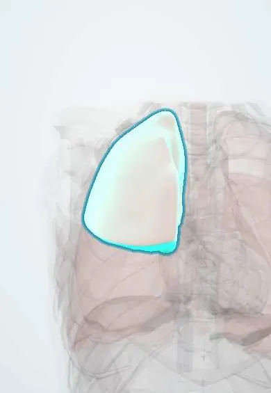
Representative model, for educational purposes only.