
Lymphangioleiomyomatosis
A rare disease of women in which abnormal smooth-muscle-like cells grow in the lungs and form many cysts, causing breathlessness and collapsed lungs.
Quick facts
- Key signs
- Progressive exertional breathlessness · Cough
- Diagnosis
- Serum VEGF-D
Red flags ↓
No doses: the clinician sets them by the current guideline.
What it is
A rare, low-grade, metastasising neoplasm of smooth-muscle-like (LAM) cells that infiltrate the lungs, lymphatics and kidneys, causing diffuse thin-walled lung cysts. It occurs sporadically or with tuberous sclerosis complex (TSC) and affects almost only women.
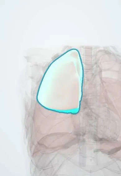
Who gets it
Sporadic LAM affects about 3 to 8 per million women, typically diagnosed in their 30s and 40s. In tuberous sclerosis complex, cysts are found on CT in about a third of adult women, often with mild disease. Very rare in men, almost always with TSC.
Causes
- Mutations of TSC2 (more often) or TSC1, which encode tuberin and hamartin, activating the mTOR pathway
- Sporadic LAM: somatic mutations in LAM cells
- TSC-LAM: germline mutation in tuberous sclerosis complex
- Oestrogen dependence: disease is linked to the reproductive years and may worsen in pregnancy or with oestrogen therapy
Risk factors
- Female sex
- Reproductive age
- Tuberous sclerosis complex
- Exogenous oestrogen and pregnancy (may accelerate the disease)
Pathophysiology
Loss of TSC2 (or TSC1) function → the tuberin-hamartin complex no longer restrains mTOR → LAM cells grow and migrate; they probably arise outside the lung (possibly uterus or lymphatics) and spread through lymphatics and blood → in the lung they release matrix-degrading enzymes and VEGF-D → alveolar walls are destroyed into many round, thin-walled cysts → airflow obstruction, a low DLCO, and pneumothorax when subpleural cysts rupture → LAM cells block the lymphatics and thoracic duct → chylous effusions and lymphangioleiomyomas. Oestrogen drives growth, so the disease belongs to the reproductive years and can worsen in pregnancy.
Classification
- Sporadic LAM versus TSC-associated LAM
- By lung function or extent of cysts: mild, moderate or severe
- Extrapulmonary: renal angiomyolipomas, lymphangioleiomyomas, chylous effusions
Symptoms and signs
- Progressive exertional breathlessness, often first misdiagnosed as asthma or COPD
- Sudden pleuritic chest pain and breathlessness from pneumothorax, often recurrent and sometimes the first sign
- Cough, wheeze and occasional haemoptysis
- Chylous pleural effusion or ascites
- Abdominal or flank pain or haematuria from bleeding angiomyolipoma
- Abdominal swelling or leg lymphoedema from lymphangioleiomyomas
Diagnosis
- Clinical
- Suspected in a woman with characteristic cysts on CT, especially with spontaneous pneumothorax, a chylous effusion or tuberous sclerosis. Examination for TSC skin signs (facial angiofibromas, hypomelanotic macules, periungual fibromas) is part of the workup.
- Tests
- Serum VEGF-D: a high level with typical CT cysts confirms LAM without biopsy. Lung function: obstructive pattern with low DLCO, sometimes with bronchodilator response. Pleural fluid with chylomicrons and high triglycerides in chylous effusions. Biopsy (transbronchial or surgical) showing LAM cells that stain for smooth-muscle actin and HMB-45 when VEGF-D and other features are not diagnostic.
- Imaging
- High-resolution CT: many round, thin-walled cysts of similar size spread evenly through both lungs, including the bases. Abdominal and pelvic CT or MRI for angiomyolipomas, lymphangioleiomyomas and lymph node enlargement; brain MRI if TSC is suspected.
Differential diagnosis
- Pulmonary Langerhans cell histiocytosis (smokers, upper-zone irregular cysts and nodules)
- Emphysema
- Birt-Hogg-Dubé syndrome (basal cysts near the pleura, skin fibrofolliculomas, renal tumours)
- Lymphocytic interstitial pneumonia (Sjögren disease)
- Asthma or COPD (common misdiagnoses)
- Light chain deposition disease and other rare cystic diseases
Thin-walled lung cysts: which disease?
| Condition | Who | Cyst pattern | Other clue |
|---|---|---|---|
| Lymphangioleiomyomatosis | Women of reproductive age; tuberous sclerosis | Round, uniform, thin-walled cysts spread evenly, bases included | Chylous effusion, renal angiomyolipoma, high VEGF-D |
| Pulmonary Langerhans cell histiocytosis | Young adult smokers | Irregular cysts with nodules, upper and middle zones; bases spared | Smoking; diabetes insipidus |
| Birt-Hogg-Dubé syndrome | Family history of cysts or kidney tumours | Basal, lens-shaped cysts beside the mediastinum and pleura | Skin fibrofolliculomas, kidney tumours |
| Lymphocytic interstitial pneumonia | Sjögren disease | A few cysts along vessels, with ground glass | Dry eyes and mouth, anti-Ro |
| Emphysema (COPD) | Older smokers | Low-attenuation areas without visible walls | Airflow obstruction, smoking history |
Goal of treatment
Slow loss of lung function with mTOR inhibition, prevent and treat pneumothorax and chylous complications, and manage angiomyolipomas.
Treatment
- Medical
- mTOR inhibitors (sirolimus, or everolimus in some patients) stabilise lung function in patients with abnormal or declining FEV1 and shrink angiomyolipomas and lymphangioleiomyomas; they are used long term because decline resumes when stopped. Inhaled bronchodilators if there is reversibility.
- Procedure
- Pleurodesis (chemical or surgical) after the first pneumothorax because recurrence is very common.
- Procedure
- Embolisation of large or bleeding renal angiomyolipomas; drainage and dietary measures for chylous effusions.
- Rehabilitation
- Pulmonary rehabilitation, supplemental oxygen, vaccination; avoidance of oestrogen-containing medicines and counselling about pregnancy and air travel.
- Surgical
- Lung transplantation for end-stage disease; LAM can recur in the graft but rarely matters clinically.
- Follow-up
- Lung function every few months at first, then yearly; periodic abdominal imaging for angiomyolipomas; side effects of mTOR inhibitors (mouth ulcers, raised lipids, infections).
Complications
- Recurrent pneumothorax
- Chylothorax and chylous ascites
- Bleeding renal angiomyolipoma
- Progressive airflow obstruction and chronic respiratory failure
- Pulmonary hypertension
Common traps
- A young woman with breathlessness and wheeze has asthma: obstruction with a pneumothorax or chylous effusion needs a CT; LAM is often labelled asthma or COPD for years
- Lung cysts always need a biopsy to prove LAM: typical CT cysts with a raised serum VEGF-D (or tuberous sclerosis, angiomyolipoma or chylous effusion) are diagnostic without one
- Pleurodesis waits for the second pneumothorax: recurrence is so common in LAM that it is offered after the first
- mTOR inhibitors cure LAM and can be stopped once stable: they stabilise lung function only while taken; decline resumes when they stop
- LAM only affects women with tuberous sclerosis: most cases are sporadic, from somatic TSC2 mutations in the LAM cells
Prognosis
Variable and usually slowly progressive: median transplant-free survival is now over 20 years from diagnosis. Disease tends to progress faster when it starts before menopause and slows after it. mTOR inhibitors have changed the outlook by stabilising lung function.
Test yourself
Think of the answer, then tap the question to see it.
Which pathway is overactive in lymphangioleiomyomatosis, and what is the targeted treatment?
mTOR, from loss of TSC1 or TSC2 function; it is treated with mTOR inhibitors such as sirolimus.
Which blood test with typical CT cysts can confirm LAM without a biopsy?
A raised serum VEGF-D (vascular endothelial growth factor D).
How do LAM cysts differ from those of pulmonary Langerhans cell histiocytosis?
LAM cysts are round, thin-walled and evenly spread including the bases; PLCH cysts are irregular, with nodules, mainly in upper zones of smokers.
Why is pleurodesis offered after the first pneumothorax in LAM?
Because pneumothorax recurs in most patients; pleurodesis reduces recurrence.
A 34-year-old woman treated for 'asthma' has had two pneumothoraces. CT: dozens of round thin-walled cysts; a fatty kidney mass. Which blood test confirms it, and what pathway is treated?
Serum VEGF-D confirms lymphangioleiomyomatosis (with angiomyolipoma). mTOR inhibitors stabilise lung function; offer pleurodesis and avoid oestrogen-containing medicines.
In dermatology
Connections
Educational information, not medical advice. For any symptom, see a health professional.
Sources: Lymphangioleiomyomatosis (StatPearls (NCBI Bookshelf), 2023); Tuberous Sclerosis Complex (StatPearls (NCBI Bookshelf), 2025).
Spotted a mistake?We are always improving for the community. Tell us what is wrong and our team will review it.Report a mistake
Representative model, for educational purposes only.