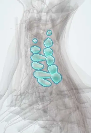
Neck facet joints !
The cervical facet joints are pairs of small sliding joints at the back of the neck; they guide nodding, turning and side bending.
Official name: Zygapophysial (facet) joints
Opens in the Durgical app, no sign-up needed.
What it is
Pairs of small sliding joints at the back of the neck, set at an angle that allows nodding, turning and side bending.
What it does
Guide and limit movement (lumbar facets block twisting), and share the load with the disc, especially when you lean back.
Key facts
- Where
- Between articular processes from C2–C3 to C7–T1 (zygapophysial joints)
- Function
- Synovial plane joints; facets about 45° to horizontal, allow flexion-extension, side bending and rotation; nerve: medial branches of dorsal rami (C2–C3: third occipital nerve)
Relations
- In front
- The intervertebral foramen with the exiting nerve root and its ganglion; further forward, the uncovertebral joint and the vertebral artery in the transverse foramina
- Behind
- Multifidus, semispinalis and the deep extensors; the medial branch of the dorsal ramus curves around the waist of the articular pillar
- Medial
- Lamina and ligamentum flavum, which blends with the front of the capsule; the spinal cord lies just inside
- Lateral
- Outer edge of the articular pillar (lateral mass), the landmark for lateral mass screws, with the vertebral artery in front
How to find it
With the person lying on their back and the neck relaxed, the articular pillars are felt as a firm column roughly two fingerbreadths to each side of the spinous processes, under the paraspinal muscles; tenderness over one level is sought when facet pain is suspected.
Examination
- Extension-rotation (quadrant) test: the neck is extended and turned to one side; local neck pain without arm symptoms suggests a facet source, while pain spreading down the arm points to root compression in the foramen (Spurling sign)
On imaging
CT shows facet osteoarthritis (joint narrowing, osteophytes, subchondral sclerosis) and fractures of the articular processes. In dislocation, the lateral view shows perched or jumped facets and axial CT shows the empty 'naked facet'. Oblique views and CT show how much an enlarged facet narrows the foramen.
Common problems
- Neck pain and headaches from arthritis or after whiplash.
- Bone spurs that narrow the opening for a nerve root.
How it is diagnosed
There is no single sign. CT or MRI show arthritis; a numbing injection of the nerves to the joint (medial branch block) shows whether the joint is the pain source.
Treatment
- Physical therapy, medicine, injections; radiofrequency ablation for lasting facet pain.
Clinical notes
- Each joint from C3–C4 down is supplied by the medial branches of two levels; the waist of the articular pillar is the fluoroscopic target for medial branch blocks and radiofrequency neurotomy
- The C2–C3 joint is supplied by the third occipital nerve; its pain is a classic cause of cervicogenic headache
- Facet hypertrophy narrows the foramen from behind while uncovertebral spurs narrow it from in front, squeezing the root between them
Related conditions
Test yourself
Think of the answer, then tap the question to see it.
Which nerves supply the cervical facet joints?
The medial branches of the dorsal rami, two levels per joint from C3–C4 down; the C2–C3 joint is supplied by the third occipital nerve.
Pain from which cervical facet joint is a classic cause of cervicogenic headache?
The C2–C3 facet joint, supplied by the third occipital nerve; upper cervical facets refer pain to the head and suboccipital region.
On a lateral X-ray, how far is the vertebral body shifted forward in a unilateral versus a bilateral cervical facet dislocation?
About a quarter of its depth in a unilateral dislocation and about half in a bilateral one.
How do unilateral and bilateral cervical facet dislocations usually present neurologically?
Unilateral ones often present with a radiculopathy; bilateral ones more often with spinal cord injury.
Educational information, not medical advice. For any symptom, see a health professional.
Its names
- Official Latin (TA2)
- articulationes zygapophysiales TA2 1707
- Official Spanish (DeCS)
- Articulación cigapofisaria DeCS D021801
- English (TA2)
- Zygapophysial (facet) joints
- Also called
- facet joints · juncturae zygapophyseales · zygapophysial joints
Related
Sources: Terminologia Anatomica (TA2); DeCS; Anatomy, Head and Neck: Cervical Spine.
Spotted a mistake?We are always improving for the community. Tell us what is wrong and our team will review it.Report a mistake
Representative model, for educational purposes only.