
Chronic progressive external ophthalmoplegia
A mitochondrial disease causing slow, symmetric droopy lids and limited eye movements, usually without double vision. Kearns-Sayre adds heart block.
What it is
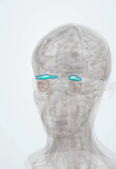
Chronic progressive external ophthalmoplegia (CPEO) is a mitochondrial myopathy with slowly worsening, usually symmetric, bilateral ptosis and ophthalmoplegia, so double vision is uncommon. Kearns-Sayre syndrome is the severe form, starting before age 20 with pigmentary retinopathy and cardiac conduction block.
Who gets it
Rare; the most common ocular presentation of mitochondrial disease. Usually begins in young adulthood; Kearns-Sayre starts before 20 years.
Causes
- Single large deletion of mitochondrial DNA (sporadic), as in Kearns-Sayre syndrome
- Point mutations of mtDNA (for example m.3243A>G, also seen in MELAS)
- Nuclear genes affecting mtDNA maintenance: POLG, TWNK, SLC25A4, RRM2B (multiple deletions, autosomal dominant or recessive)
Risk factors
- Family history of mitochondrial disease
- Maternal inheritance for point mutations
Pathophysiology
Defective oxidative phosphorylation affects tissues with high energy demand. The extraocular muscles and levator, which have very high mitochondrial content, weaken first. Muscle biopsy shows ragged-red fibres and cytochrome c oxidase-negative fibres.
Classification
- Isolated CPEO
- CPEO plus: with proximal myopathy, neuropathy, ataxia, deafness or other features
- Kearns-Sayre syndrome: onset before 20, pigmentary retinopathy and one of heart block, cerebellar ataxia or high CSF protein
Symptoms and signs
- Slowly progressive drooping of both upper lids
- Limited eye movements in all directions with little or no double vision
- Weak eye closure (orbicularis weakness)
- Exercise intolerance or limb weakness
- Hearing loss, short stature or poor night vision in syndromic forms
Diagnosis
- Clinical
- Bilateral symmetric ptosis and ophthalmoplegia with slow progression, normal pupils and no fatigability; fundus check for salt-and-pepper retinopathy.
- Tests
- Electrocardiogram and Holter for conduction block, creatine kinase, lactate, muscle biopsy (ragged-red fibres), mtDNA analysis in muscle and nuclear gene panels; antibody testing to exclude myasthenia.
- Imaging
- MRI of the orbits may show thin extraocular muscles; brain MRI in syndromic forms.
Differential diagnosis
- Myasthenia gravis
- Oculopharyngeal muscular dystrophy
- Myotonic dystrophy
- Thyroid eye disease
- Progressive supranuclear palsy (vertical gaze, but doll's eye movements preserved)
Slow bilateral ptosis and ophthalmoplegia: CPEO vs its look-alikes
| Feature | CPEO | Myasthenia gravis | Oculopharyngeal muscular dystrophy | Thyroid eye disease |
|---|---|---|---|---|
| Course | Slow over years, steady | Fluctuating, worse later in the day | Slow, usually starting in later adulthood | Active phase, then stable |
| Double vision | Rare | Common | Uncommon | Common |
| Symmetry | Symmetric | Often asymmetric | Symmetric | Often asymmetric |
| Lids | Ptosis, weak closure | Fatigable ptosis | Ptosis | Lid retraction, not ptosis |
| Swallowing | Usually normal | Can be fatigable | Dysphagia prominent | Normal |
| Key test | ECG, muscle biopsy (ragged-red fibres), mtDNA analysis | Ice-pack test, antibodies | Genetic test (PABPN1) | Thyroid tests, orbital imaging (enlarged muscles) |
Goal of treatment
Protect the cornea, improve the lid position and screen for heart and systemic complications; there is no cure.
Treatment
- Surgical
- Conservative ptosis repair (frontalis sling or limited levator surgery) to avoid corneal exposure.
- Procedure
- Pacemaker for conduction block in Kearns-Sayre syndrome.
- Conservative
- Lubricants, lid crutches on glasses and prisms for any deviation.
- Follow-up
- Regular electrocardiograms, hearing, endocrine and retinal review; genetic counselling.
Complications
- Complete heart block and sudden death (Kearns-Sayre)
- Exposure keratopathy
- Hearing loss, diabetes, hypoparathyroidism
- Retinal pigmentary changes
Common traps
- Bilateral ptosis with ophthalmoplegia is myasthenia until proved otherwise: myasthenia fluctuates, fatigues and causes diplopia, while CPEO is slow, steady, symmetric and seldom causes double vision; pupils are normal in both
- CPEO is only an eye problem: Kearns-Sayre and other forms can cause complete heart block and sudden death, so every patient needs ECG screening and follow-up
- Ptosis surgery should lift the lids fully: weak eye closure, limited upgaze and a poor Bell phenomenon make full correction risky, so repair is deliberately conservative to avoid exposure keratopathy
- Mitochondrial disease is always maternally inherited: single large mtDNA deletions are usually sporadic and nuclear genes such as POLG cause autosomal forms; only mtDNA point mutations pass down the maternal line
- Limited movement in all directions means a supranuclear palsy: in CPEO the muscles themselves are weak, so doll's-eye movements do not overcome the limit, whereas in progressive supranuclear palsy they do
Prognosis
Isolated CPEO progresses slowly with normal life expectancy; Kearns-Sayre syndrome carries cardiac and neurological risks that shorten survival.
Test yourself
Think of the answer, then tap the question to see it.
A 30-year-old has had slowly worsening ptosis and limited eye movements in all directions for years, but no double vision. Why no diplopia?
Both eyes weaken slowly and equally, so they stay roughly aligned and the brain adapts; diplopia needs misalignment.
A 16-year-old with ptosis, ophthalmoplegia and salt-and-pepper retinopathy faints. What is the likely cause and the urgent test?
Kearns-Sayre syndrome with complete heart block: urgent ECG and cardiac monitoring; a pacemaker may be needed.
Why is ptosis surgery in CPEO kept conservative?
Weak orbicularis, limited upgaze and a poor Bell phenomenon leave the cornea unprotected if the lids are lifted fully, risking exposure keratopathy.
Which bedside manoeuvre separates CPEO from progressive supranuclear palsy?
Doll's-eye (oculocephalic) movements: they overcome the limit in progressive supranuclear palsy, a supranuclear problem, but not in CPEO, where the muscles are weak.
What does a muscle biopsy show in CPEO, and why?
Ragged-red and cytochrome c oxidase-negative fibres: mitochondria proliferate under the sarcolemma to compensate for defective oxidative phosphorylation.
In neurology
Connections
Educational information, not medical advice. For any symptom, see a health professional.
Sources: Chronic Progressive External Ophthalmoplegia (CPEO) (EyeWiki (American Academy of Ophthalmology), 2025); Kearns-Sayre Syndrome (StatPearls Publishing (NCBI Bookshelf), 2023); Progressive external ophthalmoplegia: MedlinePlus Genetics (MedlinePlus, US National Library of Medicine, 2026).
Spotted a mistake?We are always improving for the community. Tell us what is wrong and our team will review it.Report a mistake
Representative model, for educational purposes only.