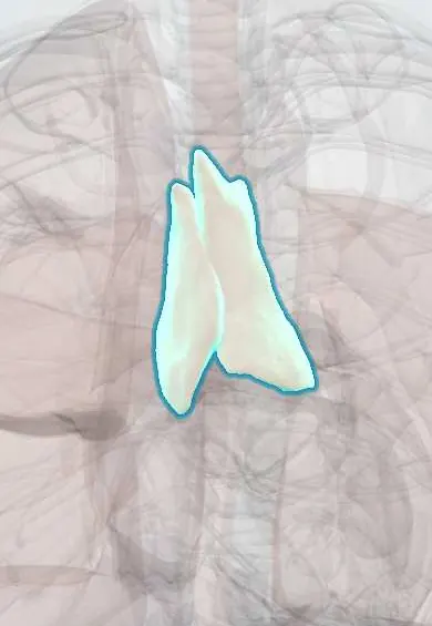
Mediastinal masses (thymoma and anterior mediastinal mass) !
Lumps in the central chest between the lungs; their site helps predict their cause, and those in front are often thymoma or lymphoma.
Updated 2026-11-01
Structures involved
Tap one to see what it is and open it in 3D.
- Thymus
- Mediastinal lymph nodes
- Trachea
- Superior vena cava
- Brachiocephalic veins
- Pericardium
- Esophagus
- Phrenic nerves
- Sympathetic trunk
- Aortic arch
Why it happens
- Benign vs malignant tumours: the comparison table
- Paraneoplastic syndromes: the table to know
- Thymus and tonsils under the microscope
What it is
Any tumour, cyst or enlarged node in the mediastinum. Location guides the differential: anterior masses are classically the «4 Ts» (thymoma, teratoma and germ cell tumours, «terrible» lymphoma, thyroid goitre); middle masses are mostly lymph nodes and cysts; posterior masses are mostly neurogenic tumours.
Who gets it
Thymoma is the most common anterior mediastinal tumour in adults, usually between 40 and 60 years. Lymphoma predominates in young adults and children, and germ cell tumours in young men. In Colombia and Latin America, tuberculosis and endemic fungal infections (histoplasmosis) are important causes of enlarged mediastinal nodes.
Causes
- Anterior: thymoma and thymic carcinoma, teratoma and other germ cell tumours, Hodgkin and non-Hodgkin lymphoma, retrosternal goitre
- Middle: lymphadenopathy (lymphoma, metastases, sarcoidosis, tuberculosis, histoplasmosis), bronchogenic and pericardial cysts
- Posterior: neurogenic tumours (schwannoma, neurofibroma, neuroblastoma in children), oesophageal lesions, aortic aneurysm
Risk factors
- Age: lymphoma and germ cell tumours in young people, thymoma in middle age
- Male sex for germ cell tumours
- Myasthenia gravis and other autoimmune diseases (thymoma)
- Tuberculosis or fungal exposure (granulomatous nodes)
- Known primary cancer (nodal metastases)
Pathophysiology
Masses cause symptoms by compressing or invading neighbouring structures in a confined space: the trachea and bronchi (cough, stridor), the superior vena cava (venous congestion), the oesophagus (dysphagia), the phrenic and recurrent laryngeal nerves and the sympathetic chain. Thymomas arise from thymic epithelial cells and disturb T-cell maturation, which explains their link with paraneoplastic autoimmune diseases such as myasthenia gravis, pure red cell aplasia and hypogammaglobulinaemia.
Classification
- By compartment: anterior (prevascular), middle (visceral), posterior (paravertebral)
- Thymic epithelial tumours by WHO histology (types A, AB, B1–B3, thymic carcinoma)
- Masaoka-Koga stage for thymoma, according to capsular and adjacent invasion
Symptoms and signs
- Many are found incidentally on a chest X-ray or CT
- Cough, chest pain or pressure, breathlessness
- Fatigable weakness, ptosis and double vision (myasthenia gravis with thymoma)
- Fever, night sweats, weight loss and pruritus (lymphoma)
- Hoarseness, dysphagia, Horner syndrome from nerve or oesophageal involvement
Diagnosis
- Clinical
- Location on cross-sectional imaging, age, sex and associated features (myasthenia, B symptoms, raised tumour markers) narrow the differential. Patients with large anterior masses need careful airway assessment before any sedation or general anaesthesia.
- Tests
- Serum AFP and beta-hCG (germ cell tumours), LDH; acetylcholine receptor antibodies (thymoma); thyroid function. Tissue diagnosis by CT-guided core biopsy, endobronchial or oesophageal ultrasound-guided needle aspiration, mediastinoscopy or surgical biopsy; a well-encapsulated thymoma may be resected without prior biopsy. Flow cytometry for lymphoma.
- Imaging
- Chest X-ray: widened mediastinum, silhouette signs. Contrast-enhanced CT defines the compartment, invasion and vessel compression; MRI separates cystic from solid lesions; PET-CT in lymphoma and staging.
Differential diagnosis
- Thymoma and thymic carcinoma
- Hodgkin and non-Hodgkin lymphoma
- Teratoma, seminoma and non-seminomatous germ cell tumours
- Retrosternal goitre
- Benign thymic hyperplasia, cysts and aortic aneurysm
Goal of treatment
Establish the diagnosis safely, protect the airway and great vessels, and treat the specific tumour with curative intent when possible.
Treatment
- Surgical
- Complete surgical resection (thymectomy) by sternotomy or minimally invasive approach is the treatment of choice for thymoma, with adjuvant radiotherapy in invasive stages; thymectomy also benefits myasthenia gravis.
- Medical
- Lymphomas and most germ cell tumours are treated primarily with chemotherapy, with or without radiotherapy, rather than surgery.
- Acute
- In airway or superior vena cava compression: upright positioning, avoidance of general anaesthesia and muscle relaxants when possible, glucocorticoids or urgent radiotherapy only after tissue is obtained when feasible, and stenting in selected cases.
- Follow-up
- Nursing: watch for stridor, positional breathlessness, facial swelling and myasthenic weakness; long-term CT follow-up after thymoma resection because of late recurrence.
Complications
- Airway compression and respiratory arrest under anaesthesia
- Superior vena cava syndrome
- Myasthenic crisis
- Pleural or pericardial spread and effusion
- Phrenic or recurrent laryngeal nerve palsy
Prognosis
Depends on the cause. Completely resected early thymomas have excellent long-term survival; thymic carcinoma and invasive stages do worse. Many mediastinal lymphomas and germ cell tumours are curable with chemotherapy.
Test yourself
Think of the answer, then tap the question to see it.
What are the «4 Ts» of an anterior mediastinal mass?
Thymoma, teratoma (germ cell tumour), «terrible» lymphoma and thyroid (retrosternal goitre).
Which autoimmune disease is classically associated with thymoma?
Myasthenia gravis; pure red cell aplasia and hypogammaglobulinaemia are other paraneoplastic associations.
Which tumour markers are measured in a young man with an anterior mediastinal mass?
AFP and beta-hCG, together with LDH, to look for germ cell tumours.
What is the most common posterior mediastinal mass?
A neurogenic tumour (schwannoma or neurofibroma in adults, neuroblastoma in young children).
What is a key anaesthetic risk of a large anterior mediastinal mass?
Collapse of the airway or great vessels after sedation, general anaesthesia or lying flat.
Related conditions
- Myasthenia gravis
- Hodgkin lymphoma
- Non-Hodgkin lymphoma
- Superior vena cava syndrome
- Goitre
- Testicular cancer
- Lung cancer
- Sarcoidosis
- Pulmonary tuberculosis
Educational information, not medical advice. For any symptom, see a health professional.
Sources: Mediastinal tumor (MedlinePlus Medical Encyclopedia (U.S. National Library of Medicine), 2024); Thymus Cancer (MedlinePlus (U.S. National Library of Medicine), 2024).
Spotted a mistake?We are always improving for the community. Tell us what is wrong and our team will review it.Report a mistake
Representative model, for educational purposes only.