
Mucormycosis
A mould infection that kills tissue within days in people with uncontrolled diabetes or weak immunity. About half die; speed of surgery and treatment decides.
What it is
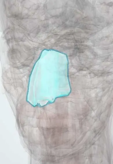
Invasive infection by moulds of the order Mucorales (Rhizopus, Mucor and related genera) that grow into blood vessels and cause rapid tissue death. The main forms are rhino-orbito-cerebral, pulmonary, cutaneous, gastrointestinal and disseminated, and the host's weakness largely decides which one appears.
Who gets it
Uncommon, but overall mortality is about 50%. Rhino-orbito-cerebral and pulmonary disease are the commonest forms. During the COVID-19 pandemic India saw a large outbreak of COVID-19-associated mucormycosis: in a multicentre study 92% of cases were rhino-orbital, linked to cumulative corticosteroid dose, diabetes and ketoacidosis, and 12-week mortality was about 32%.
Causes
- Moulds of the order Mucorales (Rhizopus, Mucor, Lichtheimia and others), found in soil and decaying organic matter
- Inhaled spores (sinuses, lungs), inoculation into wounds or burns, or ingestion
- Not spread from person to person
Risk factors
- Uncontrolled diabetes, above all diabetic ketoacidosis
- Systemic corticosteroids, including those given for severe COVID-19
- Persistent neutropenia, haematological cancer, stem cell or solid organ transplantation
- Iron overload and deferoxamine therapy
- Burns, trauma and surgical wounds; injection drug use; malnutrition and prematurity
- Previous voriconazole exposure in some patients (breakthrough infection)
Pathophysiology
Spores reach the nose, sinuses, lungs or a wound → in a host whose neutrophils are absent or crippled (ketoacidosis, high glucose, corticosteroids, neutropenia) the spores germinate unchecked → hyphae grow into small arteries and veins, causing thrombosis → the tissue downstream infarcts, so mucosa turns from red to violaceous and finally black (eschar), which is why the black eschar is a late sign of dead tissue → dead tissue has no blood supply, so antifungals cannot reach it and only surgery removes it → from the sinuses the fungus spreads to the orbit (proptosis, ophthalmoplegia, blindness), the cavernous sinus and the brain; from the lungs it spreads through the blood, most often to the brain.
Classification
- Rhino-orbito-cerebral: nose and sinuses, then orbit and brain; typical of diabetes and corticosteroid use
- Pulmonary: typical of haematological cancer and profound neutropenia
- Cutaneous: primary after burns or trauma, or secondary from the blood
- Gastrointestinal: malnourished patients and premature infants, the commonest form in neonates
- Disseminated: from any form, most often from the lungs in neutropenic patients
Symptoms and signs
- Early rhino-orbital: one-sided facial pain or swelling, nasal stuffiness, foul smell, bloody or dark nasal discharge, toothache, facial numbness
- Orbital spread: eyelid swelling, proptosis, double vision, drooping lid, sudden loss of vision
- Late: black necrotic eschar on the palate, turbinates or facial skin
- Cerebral spread: altered consciousness, cranial nerve palsies, hemiparesis
- Pulmonary: fever despite broad-spectrum antibiotics, cough, pleuritic pain, haemoptysis
Diagnosis
- Clinical
- Suspected on the host and the tempo, not on the eschar: an immunocompromised or ketoacidotic patient with one-sided facial, sinus, dental or eye symptoms, or a neutropenic patient with new lung lesions, needs urgent nasal endoscopy or imaging and biopsy the same day.
- Tests
- Tissue biopsy for histopathology and culture is the diagnosis (expert pathology is needed to tell it from other moulds). There is no routine blood test: beta-D-glucan and Aspergillus galactomannan do not detect Mucorales. Check glucose, ketones, acid-base status and neutrophil count.
- Imaging
- CT or MRI of the sinuses, orbit and brain to map the extent before surgery; chest CT in neutropenic or transplant patients with fever or respiratory symptoms.
Differential diagnosis
- Invasive aspergillosis
- Bacterial orbital cellulitis from sinusitis
- Cavernous sinus thrombosis
- Bacterial pneumonia or lung abscess in a neutropenic patient
- Sinonasal tumour or necrotising vasculitis
Black, swollen face or new lung lesions in a vulnerable host: which one?
| Condition | Typical host | Clue | Key test | Mainstay of treatment |
|---|---|---|---|---|
| Mucormycosis | Diabetic ketoacidosis, corticosteroids, iron overload or deferoxamine, transplant, neutropenia | Rapid necrosis; black eschar comes late; may break through voriconazole | Biopsy (histology and culture); galactomannan and beta-D-glucan are negative | Emergency surgery plus amphotericin B, and correct the host factor |
| Invasive aspergillosis | Prolonged neutropenia, transplant, high-dose corticosteroids, severe influenza or COVID-19 in intensive care | Lung nodules; sinus disease less common than in mucormycosis | Galactomannan in serum or lavage supports it; biopsy | Voriconazole or another mould-active azole |
| Bacterial orbital cellulitis | Often a child or adult with acute sinusitis; no immune defect needed | Fever, painful eye movements, proptosis, no necrotic mucosa | Orbital CT; cultures | Intravenous antibiotics, drainage of abscess when present |
Goal of treatment
Within hours of suspicion: remove all dead tissue, start an active antifungal and reverse what weakened the host, because survival falls with every day of delay.
Treatment
- Surgical
- Emergency surgical debridement of all necrotic tissue, repeated as needed (sinus surgery, resection of palate or lung, and orbital exenteration when the eye is lost and the brain is threatened), decided by a multidisciplinary team.
- Medical
- Antifungal started at once, before culture results: lipid amphotericin B is first line; isavuconazole or posaconazole are alternatives or step-down options, as the guideline sets. Voriconazole has no activity against Mucorales and is never used for it.
- Acute
- Correct the host factor: treat diabetic ketoacidosis and control glucose, reduce or stop corticosteroids and other immunosuppressants where possible, stop deferoxamine, and support neutrophil recovery.
- Follow-up
- Repeated endoscopy and imaging to look for residual necrosis, and monitoring of kidney function and electrolytes during amphotericin B therapy.
Complications
- Blindness and loss of the eye
- Cavernous sinus and internal carotid thrombosis, stroke and brain abscess
- Facial and palatal disfigurement after debridement
- Massive haemoptysis in pulmonary disease
- Dissemination and death
Common traps
- No black eschar means no mucormycosis: the eschar is dead tissue and comes late; facial pain, swelling, numbness or eye signs in a ketoacidotic or immunocompromised patient are enough to act
- A broad antifungal such as voriconazole covers any mould: voriconazole treats Aspergillus but has no activity against Mucorales, and mucormycosis can break through voriconazole prophylaxis
- A negative galactomannan or beta-D-glucan makes an invasive mould unlikely: neither test detects Mucorales, so biopsy decides
- Amphotericin B alone can cure it: dead tissue has no blood flow for the drug to reach, so emergency debridement is required
- Once the antifungal is running the underlying disease can wait: uncorrected ketoacidosis, high-dose corticosteroids or neutropenia let the fungus keep advancing
Prognosis
Overall mortality is about 50%; it is higher with brain involvement, dissemination, persistent neutropenia and delayed diagnosis. Early diagnosis, surgery and an active antifungal improve survival.
Prevention
- Good glucose control and prompt treatment of ketoacidosis
- Corticosteroids only when indicated, at the lowest effective intensity, with glucose monitoring (lessons from COVID-19)
- Mould-active prophylaxis in selected high-risk patients, as the guideline sets
- Wound care for burns and soil-contaminated injuries
Test yourself
Think of the answer, then tap the question to see it.
Why does mucormycosis need surgery as well as an antifungal?
The fungus invades and clots blood vessels, so the infected tissue dies; dead tissue has no blood supply for the drug to reach, and only debridement removes it.
Which host factors steer mucormycosis to the sinuses rather than the lungs?
Uncontrolled diabetes, especially ketoacidosis, and corticosteroids typically cause rhino-orbito-cerebral disease; profound neutropenia and haematological cancer typically cause pulmonary disease.
A man with diabetic ketoacidosis has one-sided facial pain, eyelid swelling and a stuffy nose, but no eschar. What do you do?
Treat it as possible mucormycosis now: urgent endoscopy, imaging and biopsy, start amphotericin B, call the surgical team and correct the ketoacidosis. The eschar is a late sign, not a requirement.
A neutropenic patient on voriconazole prophylaxis develops new lung nodules. Which mould must you think of, and why?
Mucorales: voriconazole controls Aspergillus but not Mucorales, so mucormycosis can break through it; galactomannan stays negative, and a biopsy or culture is needed.
How does mucormycosis differ from invasive aspergillosis in the tests and the first drug?
Aspergillosis often has a positive galactomannan and is treated first with voriconazole; mucormycosis is galactomannan and beta-D-glucan negative and needs amphotericin B plus surgery.
Why it happens
Connections
Microorganisms that cause it1
Educational information, not medical advice. For any symptom, see a health professional.
Sources: Clinical Overview of Mucormycosis (US Centers for Disease Control and Prevention (CDC), 2025); Recognizing the Early Risk-Based Clinical Manifestations of Mucormycosis: Cornerstones for Improved Survival and Therapeutic Outcomes (Journal of Fungi (open access, PMC), 2025); Multicenter Case–Control Study of COVID-19–Associated Mucormycosis Outbreak, India (Emerging Infectious Diseases (CDC), 2023); Code Mucor: Guidelines for the Diagnosis, Staging and Management of Rhino-Orbito-Cerebral Mucormycosis in the Setting of COVID-19 (Indian Journal of Ophthalmology (PMC), 2021).
Spotted a mistake?We are always improving for the community. Tell us what is wrong and our team will review it.Report a mistake
Representative model, for educational purposes only.