
Fungal sinusitis (invasive and noninvasive)
Fungal infection of the sinuses. In diabetics with ketoacidosis or very weak immunity, mucormycosis can destroy the face within hours: an emergency.
What it is
Fungal rhinosinusitis is divided by whether hyphae invade tissue. Invasive forms, especially acute invasive mucormycosis in diabetic ketoacidosis or neutropenia, spread through vessels to the orbit and brain and are surgical emergencies; noninvasive forms (fungal ball, allergic fungal rhinosinusitis) affect immunocompetent people.
Who gets it
Acute invasive disease occurs almost only in immunocompromised people: uncontrolled diabetes (especially ketoacidosis), hematologic cancer with neutropenia, transplantation and high-dose corticosteroids. Mucormycosis surged during COVID-19 in India. Allergic fungal rhinosinusitis is more common in warm humid regions and in young atopic adults. The Mucorales organism page in the microbiology section gives the microbiology.
Causes
- Acute invasive (fulminant): Mucorales (Rhizopus, Mucor, Lichtheimia) in diabetic ketoacidosis; Aspergillus (mostly fumigatus) in neutropenia
- Chronic invasive: Aspergillus and other molds, slow course over more than 12 weeks, mild immunosuppression or diabetes
- Granulomatous invasive: Aspergillus flavus in immunocompetent hosts, mainly in Sudan, India and Pakistan
- Fungal ball (mycetoma): Aspergillus mass in one sinus, usually the maxillary, often after dental root filling material enters the sinus
- Allergic fungal rhinosinusitis: IgE-mediated hypersensitivity to dematiaceous fungi or Aspergillus in atopic patients
Risk factors
- Diabetic ketoacidosis or poorly controlled diabetes (acidosis and high free iron favor Mucorales)
- Prolonged neutropenia, leukemia, stem cell or solid organ transplantation
- High-dose or prolonged systemic corticosteroids
- Iron overload and deferoxamine therapy
- Advanced HIV infection
- Atopy, asthma and nasal polyps (allergic fungal rhinosinusitis)
- Endodontic treatment of upper molars (fungal ball)
Pathophysiology
Mucorales are angioinvasive: hyphae invade arterial walls, causing thrombosis, ischemic necrosis and black eschar. Ketoacidosis impairs neutrophil function and releases iron from transferrin, which the fungus uses to grow. From the nasal cavity and turbinates the infection spreads to the ethmoid cells, through the thin lamina papyracea to the orbit and orbital apex, to the cavernous sinus and internal carotid artery, and through the cribriform plate to the brain. Allergic fungal rhinosinusitis is a type I and III hypersensitivity response that fills sinuses with eosinophilic mucin and polyps and slowly expands and thins bone.
Classification
- Invasive: acute invasive (less than 4 weeks), chronic invasive (more than 12 weeks), granulomatous invasive
- Noninvasive: saprophytic fungal infestation (crusts), fungal ball, allergic fungal rhinosinusitis
- Allergic fungal rhinosinusitis is diagnosed with the Bent and Kuhn criteria: type I hypersensitivity, nasal polyps, characteristic CT, eosinophilic mucin without invasion, positive fungal stain
Symptoms and signs
- Acute invasive: fever, facial pain and swelling, nasal congestion with dark or bloody discharge in an immunocompromised patient
- Black or pale (ischemic) necrotic areas on the turbinates, septum or palate
- Facial numbness and loss of sensation of the cheek or nose
- Periorbital swelling, proptosis, ophthalmoplegia, ptosis, visual loss (orbital apex syndrome)
- Altered mental status, cranial nerve palsies, hemiparesis (cavernous sinus or carotid involvement)
- Fungal ball: unilateral chronic sinusitis symptoms or none
- Allergic fungal rhinosinusitis: nasal polyps, thick tan to brown mucin, sometimes proptosis or telecanthus in young patients
Diagnosis
- Clinical
- High suspicion in any immunocompromised patient with sinus symptoms: urgent nasal endoscopy looking for pale, insensitive or black mucosa (middle turbinate is the earliest site) and examination of palate, eyes and cranial nerves.
- Tests
- Urgent endoscopic biopsy with frozen section and fungal stains (calcofluor white, Gomori methenamine silver, PAS) showing tissue invasion is diagnostic: Mucorales have broad, ribbon-like, pauciseptate hyphae branching at right angles; Aspergillus has narrow, septate hyphae branching at acute angles (about 45 degrees). Cultures identify the species. Galactomannan helps in aspergillosis but not in mucormycosis. Check glucose, ketones, acid-base status and neutrophil count. In allergic fungal rhinosinusitis, total IgE is high and eosinophilic mucin with Charcot-Leyden crystals is found.
- Imaging
- Contrast CT of the sinuses shows mucosal thickening, bone erosion and soft-tissue stranding in the fat around the maxillary sinus; MRI with contrast is better for orbital, cavernous sinus and intracranial spread (non-enhancing necrotic mucosa, the 'black turbinate' sign). Fungal ball: dense or calcified spots inside an opacified sinus. Allergic fungal rhinosinusitis: expanded sinuses with heterogeneous high-density content on CT.
Differential diagnosis
- Bacterial acute or chronic rhinosinusitis
- Orbital cellulitis from bacterial sinusitis
- Cavernous sinus thrombosis
- Sinonasal malignancy, including NK/T-cell lymphoma
- Granulomatosis with polyangiitis
- Chronic rhinosinusitis with nasal polyps (versus allergic fungal rhinosinusitis)
Goal of treatment
For invasive disease, act within hours: reverse immunosuppression, give antifungals and remove all necrotic tissue. For noninvasive disease, clear the sinus and control inflammation.
Treatment
- Acute
- Emergency: admit, call otolaryngology urgently and correct the predisposing factor (treat diabetic ketoacidosis, reduce corticosteroids or immunosuppressants when possible, support neutrophil recovery).
- Medical
- Start intravenous liposomal amphotericin B as soon as mucormycosis is suspected, without waiting for culture; isavuconazole or posaconazole are alternatives or step-down options. Voriconazole is first line for invasive aspergillosis but is not active against Mucorales.
- Surgical
- Early and repeated surgical debridement of all necrotic tissue until bleeding healthy margins, endoscopic or open (maxillectomy, palate resection); orbital exenteration may be needed when the eye is lost and infection threatens the brain.
- Medical
- Chronic invasive disease: surgical debridement plus prolonged systemic antifungal therapy.
- Surgical
- Fungal ball: endoscopic sinus surgery removing the fungal mass is curative; antifungals are usually not needed.
- Medical
- Allergic fungal rhinosinusitis: endoscopic sinus surgery to remove mucin and polyps, followed by intranasal corticosteroids, saline irrigation and short courses of systemic corticosteroids; long-term follow-up because it often recurs.
Complications
- Orbital apex syndrome and blindness
- Cavernous sinus thrombosis and internal carotid artery thrombosis with stroke
- Brain abscess and intracranial spread
- Facial and palatal destruction after debridement
- Death
- Allergic fungal rhinosinusitis: bone remodelling, proptosis, recurrence
Prognosis
Acute invasive mucormycosis has a mortality of about 30 to 50%, higher with intracranial spread, and survival depends on early diagnosis, prompt surgery and recovery of immunity. Fungal ball and allergic fungal rhinosinusitis are not life-threatening.
Test yourself
Think of the answer, then tap the question to see it.
A patient in diabetic ketoacidosis develops facial pain, black necrotic turbinate and periorbital swelling. Diagnosis and first steps?
Rhino-orbito-cerebral mucormycosis: correct ketoacidosis, start IV liposomal amphotericin B and perform urgent surgical debridement.
How do Mucorales and Aspergillus hyphae differ on histology?
Mucorales: broad, ribbon-like, pauciseptate, right-angle branching; Aspergillus: narrow, septate, acute-angle branching.
Why should voriconazole not be used empirically when mucormycosis is suspected?
It has no activity against Mucorales.
An immunocompetent patient has a unilateral opacified maxillary sinus with dense calcifications after dental root canal treatment. Treatment?
Fungal ball: endoscopic removal through sinus surgery; antifungals not needed.
What are the major Bent and Kuhn criteria for allergic fungal rhinosinusitis?
Type I hypersensitivity, nasal polyps, characteristic CT findings, eosinophilic mucin without invasion, and positive fungal stain.
In ENT
- Granulomatous diseases of the nose: leishmaniasis, leprosy, TB, syphilis, rhinoscleroma, rhinosporidiosis, GPA
- Complications of sinusitis: orbital, intracranial and bony
Connections
Microorganisms that cause it1
Educational information, not medical advice. For any symptom, see a health professional.
Sources: Fungal Sinusitis (StatPearls (NCBI Bookshelf), 2026); Mucormycosis (StatPearls (NCBI Bookshelf), 2026); Rhinocerebral Mucormycosis (StatPearls (NCBI Bookshelf), 2025).
Spotted a mistake?We are always improving for the community. Tell us what is wrong and our team will review it.Report a mistake
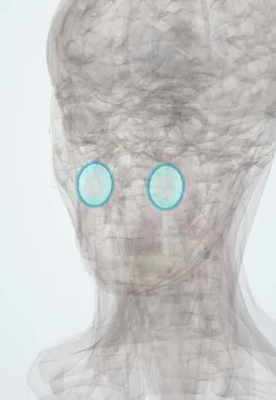
Representative model, for educational purposes only.