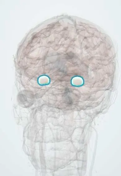
Nervous system · Head and neck
Amygdala !
The amygdala is an almond-shaped cluster of cells in the front of the temporal lobe; it processes fear and emotional memories.
Official name: Amygdaloid body
Opens in the Durgical app, no sign-up needed.
What it is
An almond-shaped nucleus in the front of the temporal lobe.
What it does
Processes fear and emotional memories.
Key facts
- Where
- Anterior temporal lobe, in front of the hippocampus
- Blood supply
- Anterior choroidal artery, MCA
- Function
- Fear and emotional memory
Relations
- Behind
- Head of the hippocampus and the tip of the temporal horn of the lateral ventricles
- Above
- Basal forebrain and the lentiform nucleus (putamen and globus pallidus)
- Medial
- Uncus, which covers it, facing the tentorial notch, the midbrain and the oculomotor nerve
- Course
- Outputs run in the stria terminalis, which arches along the caudate nucleus in the wall of the lateral ventricles to the hypothalamus, and in the ventral amygdalofugal pathway
How to find it
On coronal MRI it is a rounded grey mass in the medial temporal lobe, above and in front of the tip of the temporal horn; on sagittal images it lies directly in front of the head of the hippocampus, beneath the uncus.
On imaging
Coronal MRI, cut at right angles to the hippocampus, shows it best, above and in front of the tip of the temporal horn. In herpes simplex encephalitis, T2 and FLAIR show bright swelling of the medial temporal lobe including the amygdala, often worse on one side, and DWI may change early. In temporal lobe epilepsy, MRI looks mainly for hippocampal sclerosis. CT is insensitive.
Clinical notes
- Features of Klüver–Bucy syndrome can also appear in frontotemporal dementia and after bilateral temporal surgery
- Research links heightened amygdala responses to anxiety disorders and post-traumatic stress disorder
- It is removed with the hippocampus in selective amygdalohippocampectomy for mesial temporal lobe epilepsy
Related conditions
- Autoimmune encephalitis (including anti-NMDA receptor encephalitis)
- Major depressive disorder
- Suicidal behaviour and suicide risk
- Bipolar disorder
- Generalized anxiety disorder
- Panic disorder
- Post-traumatic stress disorder (PTSD) and acute stress disorder
- Autism spectrum disorder (ASD)
- Personality disorders (clusters A, B and C)
- Social anxiety disorder and specific phobia
- Adjustment disorder
- Conduct disorder and oppositional defiant disorder
- Intimate partner and sexual violence (mental-health care)
- Separation anxiety disorder
- Disruptive mood dysregulation disorder (DMDD)
- Prolonged grief disorder
- Epilepsy and seizures
- Alzheimer disease
Test yourself
Think of the answer, then tap the question to see it.
What is the main role of the amygdala?
It detects threat and drives the fear response through the hypothalamus and brainstem, and gives emotional weight to memories.
What is Klüver–Bucy syndrome, and what causes it?
Loss of fear, hyperorality, hypersexuality and visual agnosia, from bilateral amygdala damage, for example after herpes simplex encephalitis or trauma.
Where is the amygdala relative to the hippocampus?
In the front of the medial temporal lobe, directly in front of the head of the hippocampus and the tip of the temporal horn, beneath the uncus.
Educational information, not medical advice. For any symptom, see a health professional.
Its names
- Official Latin (TA2)
- corpus amygdaloideum TA2 5549
- Official Spanish (DeCS)
- Cuerpo amigdalino
- English (TA2)
- Amygdaloid body
- Also called
- amygdaloid complex · nucleus amygdalae
Related
Sources: Terminologia Anatomica (TA2); Neuroanatomy, Amygdala.
Spotted a mistake?We are always improving for the community. Tell us what is wrong and our team will review it.Report a mistake
Representative model, for educational purposes only.