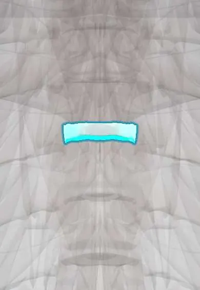
T4–T5 disc !
The T4-T5 disc is the thin cushion between the T4 and T5 vertebrae in the mid-back; it allows the small movement the ribs permit.
Official name: T4-T5
Opens in the Durgical app, no sign-up needed.
What it is
The cushion between T4 and T5: a tough outer ring of fibres (annulus fibrosus) around a soft centre.
What it does
Absorbs load and allows the small amount of movement the rib cage permits.
Key facts
- Where
- Between T4 and T5 bodies; thin thoracic disc; head of rib 5 joins it
- Attaches
- Annulus fibrosus anchored to the endplates and ring apophyses of T4 and T5; anterior and posterior longitudinal ligaments
- Function
- Absorbs and spreads load; herniation rare, often central onto the cord; lateral herniation may irritate the T4 root
Relations
- In front
- Esophagus with the tracheal bifurcation in front of it; the thoracic duct crosses from right to left behind the esophagus at about this height (texts vary: T4–T6)
- Lateral
- Head of rib 5 and its costovertebral joint; on the right the azygos vein arches forward over the right main bronchus, on the left the aortic arch becomes the thoracic aorta
- Behind
- Posterior longitudinal ligament and the spinal cord in the narrowest part of the canal
How to find it
From the front, the sternal angle (the ridge where the manubrium meets the body of the sternum, beside the 2nd costal cartilages) points straight back to this disc; from behind it lies near the tip of the T4 spinous process.
Examination
- Sensory level: pinprick is tested from the feet upward until the feeling changes; a level at the nipples suggests cord compression near T4, though the true lesion can lie higher
On imaging
On a sagittal chest CT reformat, a line drawn backward from the sternal angle reaches this disc (some texts place it at the lower border of T4). The carina and the arch of the aorta on axial slices confirm the height.
Common problems
- Thoracic disc herniations are uncommon; many are found by chance on MRI and cause no symptoms.
- A large one can press on the spinal cord, or cause band-like pain around the chest.
How it is diagnosed
MRI; CT shows whether the herniation is calcified (hardened), which matters for surgery.
Treatment
- Usually physical therapy and pain medicine; surgery only if the spinal cord is pressed.
Clinical notes
- For a transthoracic approach at this height many surgeons enter through the right chest, since the aortic arch and the heart crowd the left side
- A new sensory level at the nipples with back pain, in a person with cancer, raises concern for metastatic cord compression and calls for urgent MRI of the whole spine
Test yourself
Think of the answer, then tap the question to see it.
Which plane lies at the T4–T5 disc, and what is found at this level?
The sternal angle plane: the carina, both ends of the aortic arch and the azygos vein entering the superior vena cava; it divides the superior and inferior mediastinum.
Which root leaves at the T4–T5 disc, and where is its dermatome?
The T4 root; its dermatome is the band at the nipples, a classic landmark for a sensory level.
How is a sensory level tested in suspected cord compression near the T4–T5 disc?
Pinprick is tested from the feet upward until the feeling changes; a level at the nipples suggests compression near T4, though the true lesion can lie higher.
Educational information, not medical advice. For any symptom, see a health professional.
Its names
- Official Latin (TA2)
- discus intervertebralis T4-T5 TA2 1684
- Official Spanish (DeCS)
- Disco T4-T5 DeCS D007403
- English (TA2)
- T4-T5
- Also called
- fibrous ring · fibrocartilago intervertebralis
Related
Sources: Terminologia Anatomica (TA2); DeCS; Anatomy, Back, Intervertebral Discs; 7.3 The Vertebral Column (OpenStax (Rice University), 2022) · adapted, CC BY 4.0.
Spotted a mistake?We are always improving for the community. Tell us what is wrong and our team will review it.Report a mistake
Representative model, for educational purposes only.