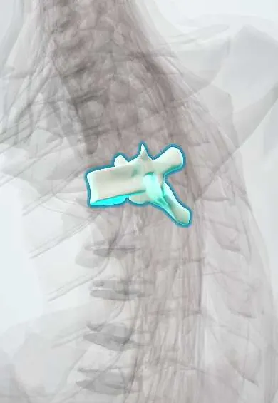
T4 vertebra !
T4 is the fourth thoracic (mid-back) vertebra; it has joints for the ribs and helps form the cage that protects the heart and lungs.
Official name: T4
Opens in the Durgical app, no sign-up needed.
What it is
The T4 mid-back vertebra, with joints on each side for the ribs.
What it does
Holds up the trunk and, with the ribs and breastbone, forms the rib cage, which protects the heart and lungs. The ribs make this part of the spine stiff.
Key facts
- Joints with
- T3 above and T5 below (discs, facet joints); heads of ribs 4 and 5; tubercle of rib 4 (costotransverse joint)
- Landmarks
- Typical thoracic: heart-shaped body; costal demifacets above and below; transverse costal facets; long sloping spine; sternal angle at T4–T5 disc (tracheal bifurcation)
Relations
- In front
- Esophagus, indented by the aortic arch; in front of it the tracheal bifurcation (carina)
- Lateral
- On the right the azygos vein arches forward over the root of the right lung; on the left the arch of the aorta becomes the descending thoracic aorta
- Behind
- Trapezius, rhomboids and erector spinae over long, downward-sloping spines
- Contents
- Spinal cord in the narrowest part of the canal (about T4–T9), with little room around it
How to find it
In front, the sternal angle (angle of Louis) is felt as a ridge where the manubrium meets the body of the sternum, with the second costal cartilages beside it; the horizontal plane through it meets the back at the T4–T5 disc. Behind, the T4 spine lies a little below the line joining the roots of the scapular spines.
On imaging
On a chest X-ray the aortic knob and the carina project near T4–T5. On an AP spine film a missing pedicle ('winking owl' sign) is a classic sign of metastatic destruction.
Common problems
- Compression fractures from osteoporosis, sometimes after a minor fall or even a cough.
- A rounded upper back (kyphosis) from posture, wedge fractures or Scheuermann disease in teens.
- Scoliosis: a sideways curve with rotation, often starting in adolescence.
How it is diagnosed
X-rays for fractures and curves (scoliosis is measured with the Cobb angle). MRI tells new fractures from old ones. A bone density scan (DEXA) checks for osteoporosis.
Treatment
- Osteoporosis fractures: pain control, a short time in a brace, and treatment of the osteoporosis.
- Painful fractures that do not settle: kyphoplasty or vertebroplasty (cement injected into the bone).
- Large or progressive curves: bracing in growing children, sometimes fusion surgery.
Clinical notes
- The thoracic spine is the commonest site of spinal metastases (breast, lung, prostate, kidney, myeloma); the pedicle and back of the body are often hit first
- Herpes zoster or a thoracic root lesion at T4 gives a band of pain or rash around the chest at nipple level
- A widened angle of the carina on a chest X-ray can point to an enlarged left atrium or to enlarged nodes beneath the carina
- Pedicles are narrowest in the upper and mid thoracic spine (about T4–T6), so pedicle screws here are planned on CT and a screw that breaches medially reaches the cord
Memory aid
Sternal angle plane, 'RATPLANT': Rib 2, Aortic arch ends, Trachea divides, Pulmonary trunk divides, Ligamentum arteriosum, Azygos joins SVC, Nerve (left recurrent laryngeal) hooks, Thoracic duct crosses.
Test yourself
Think of the answer, then tap the question to see it.
Which plane meets the back at the T4–T5 disc, and which mediastinal parts does it separate?
The sternal angle plane (angle of Louis); it separates the superior from the inferior mediastinum.
Why can an aneurysm of the aortic arch at the T4 vertebra level cause hoarseness?
The left recurrent laryngeal nerve hooks under the aortic arch at this level.
What does the «winking owl» sign on an AP film of the thoracic spine, such as at T4, indicate?
A missing pedicle, a classic sign of metastatic destruction; the thoracic spine is the commonest site of spinal metastases.
Which dermatome crosses the nipple in men?
T4, a standard landmark for sensory levels.
Educational information, not medical advice. For any symptom, see a health professional.
Its names
- Official Latin (TA2)
- vertebra thoracica IV TA2 1059
- Official Spanish (DeCS)
- Vértebra dorsal D4 DeCS D013904
- English (TA2)
- T4
Related
- T3–T4 disc
- T4–T5 disc
- Posterior intercostal arteries
- Anterior longitudinal ligament
- Rib-to-spine ligaments
- Endplates
- Mid-back facet joints
- Ligamentum flavum
Sources: Terminologia Anatomica (TA2); DeCS; Anatomy, Back, Thoracic Vertebrae.
Spotted a mistake?We are always improving for the community. Tell us what is wrong and our team will review it.Report a mistake
Representative model, for educational purposes only.