Conditions · Hematology and oncology
Carcinoma of unknown primary !
A cancer that has already spread when it is found, but whose starting organ cannot be identified even after careful testing.
Updated 2026-11-01
Structures involved
Tap one to see what it is and open it in 3D.
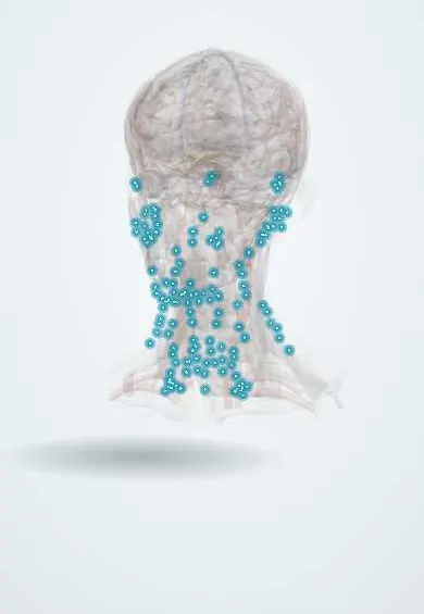
- Neck lymph nodes
- Liver
- Palatine tonsils
- Para-aortic lymph nodes
- Peritoneum
- Pancreas
- Mediastinal lymph nodes
Why it happens
- Pathways of spread and the usual metastatic sites
- Tumour markers: serum and tissue, and what each one is for
- Immunohistochemistry: the principle and the markers to know
- Invasion and metastasis: the metastatic cascade, step by step
- Differentiation and anaplasia: how a tumour cell looks under the microscope
What it is
A biopsy-proven metastatic cancer for which the primary site is not found after a standard work-up of history, examination, laboratory tests, imaging and pathology. Most are adenocarcinomas or poorly differentiated carcinomas; the teaching point is a focused work-up that looks for treatable cancers and for 'favourable subsets' that behave like a known primary.
Who gets it
It once made up several per cent of all cancers; with better imaging and pathology, fewer than 1–2% of new cancers now remain truly unknown. It is mostly a disease of older adults. In Latin America, limited access to PET-CT, immunohistochemistry panels and molecular profiling outside major centres means more patients are labelled as unknown primary and diagnosis takes longer.
Causes
- A primary tumour that is too small to detect, has regressed, or was removed before
- Early spread from the primary with a biologically aggressive phenotype
- Common hidden primaries include lung, pancreas, biliary tract, stomach and colon; less often breast, ovary, prostate and kidney
Risk factors
- Older age
- Smoking
- Risk factors of the common occult primaries (alcohol, obesity, family history)
Pathophysiology
The metastatic deposits dominate the picture while the primary remains silent. Cancers of unknown primary tend to have early and unpredictable spread to the liver, lungs, bones and lymph nodes. Immunohistochemistry reads the 'identity' of the tumour cells: cytokeratin patterns (for example CK7 and CK20) and lineage markers such as TTF-1 (lung, thyroid), CDX2 (bowel), GATA3 (breast, urothelium), PAX8 (kidney, ovary) and PSA (prostate) point to the likely origin.
Classification
- By histology: adenocarcinoma (well or moderately differentiated), poorly differentiated carcinoma, squamous cell carcinoma, neuroendocrine carcinoma, undifferentiated neoplasm
- Favourable subsets (about 20%): women with adenocarcinoma in axillary nodes (breast-like), women with serous peritoneal carcinomatosis (ovarian-like), squamous carcinoma in upper or mid cervical nodes (head and neck-like), men with osteoblastic bone metastases and high PSA (prostate-like), young men with midline tumours and high beta-hCG or AFP (germ cell-like), neuroendocrine tumours, single small metastasis
- Unfavourable subset: the rest, such as multiple liver, lung or bone metastases from adenocarcinoma
Symptoms and signs
- Symptoms from the site of metastasis: a lump in the neck, armpit or groin
- Right upper abdominal pain, jaundice or swelling from liver metastases
- Bone pain or fracture
- Cough or breathlessness from lung or pleural spread
- Abdominal distension from ascites
- Weight loss and fatigue
Diagnosis
- Clinical
- Full history and examination including breasts, pelvis, rectum, prostate, testes, skin and head and neck (with endoscopy for neck nodes). The aim is not to search endlessly but to find treatable primaries and favourable subsets quickly.
- Tests
- Core or excisional biopsy with an immunohistochemistry panel guided by the morphology; molecular profiling for actionable alterations (such as MSI-high) and, in some centres, tissue-of-origin gene expression tests. Blood count, liver and kidney function, calcium; PSA in men, beta-hCG and AFP in young men with midline disease, and CA-125 in women with peritoneal disease. Tumour markers are not used as a broad screen.
- Imaging
- CT of chest, abdomen and pelvis; mammography and breast MRI in women with axillary nodes; PET-CT especially for squamous cervical node presentations; further imaging directed by symptoms.
Differential diagnosis
- Metastatic cancer from a primary missed by an incomplete work-up
- Lymphoma presenting as an undifferentiated tumour
- Melanoma with regressed primary
- Sarcoma or germ cell tumour
- Benign causes of lymphadenopathy or liver lesions (tuberculosis, abscess)
Goal of treatment
Treat favourable subsets as their likely primary cancer, sometimes with curative intent, and for the rest give effective, well-tolerated palliative therapy while controlling symptoms.
Treatment
- Medical
- Favourable subsets are treated with the guidelines for their likely primary: for example breast cancer treatment for axillary adenocarcinoma in a woman, ovarian cancer treatment for peritoneal carcinomatosis, androgen deprivation for a prostate-like picture, and cisplatin-based chemotherapy for germ cell-like disease.
- Procedure
- Squamous cancer in neck nodes is treated like head and neck cancer with neck dissection and/or radiotherapy; a single metastasis may be removed or irradiated.
- Medical
- Unfavourable subsets: platinum-based doublet chemotherapy with palliative intent, targeted therapy or immune checkpoint inhibitors when a matching molecular marker is found.
- Conservative
- Early palliative care for pain, symptom control and support; honest communication about the uncertainty of the diagnosis.
Complications
- Spinal cord compression and pathological fractures
- Hypercalcaemia of malignancy
- Malignant effusions and ascites
- Liver failure from extensive metastases
- Venous thromboembolism
Prognosis
Poor overall, with a median survival of under a year for unfavourable subsets. Favourable subsets, single-site disease, nodal or neuroendocrine presentations do much better and some are curable; liver or adrenal spread, poor performance status, high LDH and older age predict worse outcomes.
Test yourself
Think of the answer, then tap the question to see it.
A woman has adenocarcinoma in an axillary node and no primary found. How is she treated?
As stage II–III breast cancer, after mammography and breast MRI.
Squamous carcinoma in an upper neck node with no known primary suggests which origin?
A head and neck primary, such as the tonsil or base of tongue.
Which laboratory test helps in a man with osteoblastic bone metastases of unknown primary?
Serum PSA, to identify prostate cancer.
What is the role of immunohistochemistry in carcinoma of unknown primary?
Markers such as CK7, CK20, TTF-1 and CDX2 suggest the likely tissue of origin.
Related conditions
- Head and neck squamous cell carcinoma (oral cavity and oropharynx, HPV)
- Breast cancer
- Ovarian cancer
- Prostate cancer
- Lung cancer
- Pancreatic cancer
- Testicular cancer
- Melanoma
- Cervical lymphadenopathy
- Hypercalcaemia of malignancy
- Malignant spinal cord compression
- Paraneoplastic syndromes
Educational information, not medical advice. For any symptom, see a health professional.
Sources: Cancer of Unknown Primary (CUP) Treatment (PDQ®)–Health Professional Version (National Cancer Institute, 2025).
Spotted a mistake?We are always improving for the community. Tell us what is wrong and our team will review it.Report a mistake
Representative model, for educational purposes only.