Cervical radiculopathy !
Cervical radiculopathy is a pinched nerve root in the neck; it causes pain, numbness or weakness that runs down the arm.
Updated 2026-10-29
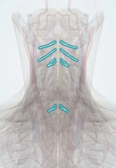
Structures involved
Tap one to see what it is and open it in 3D.
- Neck nerve roots
- C6 nerve root
- C7 nerve root
- C5 nerve root
- C5–C6 disc
- C6–C7 disc
- Neck facet joints
- Brachial plexus roots
In the clinical exam
- Deep tendon reflexes and their grading
- Sensory examination: modalities and dermatomes
- Upper vs lower motor neuron syndrome
In physiology
Why it happens
- Pain and temperature: pathways and modulation
- Peripheral nerve: endoneurium, perineurium, epineurium; myelin
What it is
Dysfunction of a cervical nerve root, usually from compression where it leaves the spine, causing neck and arm pain that follows a dermatome, with numbness, weakness or reduced reflexes in that root's territory.
Who gets it
Most common in middle age, peaking around the fifth decade. The C7 root is affected most often, followed by C6. A frequent reason for referral to spine, neurology and physiatry clinics in Colombia.
Causes
- Foraminal narrowing from degenerative change: uncovertebral and facet osteophytes, disc height loss (the most common cause, especially in older adults)
- Soft disc herniation (more typical in younger adults)
- Less often: trauma, tumour, infection or inflammatory disease
Risk factors
- Age over 40 and cervical spondylosis
- Heavy manual labour, lifting or vibration exposure
- Smoking
- Previous neck injury
- Prior cervical fusion at an adjacent level
Pathophysiology
A herniated disc or osteophytes narrow the intervertebral foramen and press on the root. Mechanical compression plus local inflammatory mediators produce root ischaemia and oedema, so pain and sensory, motor and reflex changes appear in that root's distribution. Each cervical root exits above the vertebra of the same number, so a C5–C6 disc usually affects the C6 root.
Classification
- C5: lateral arm numbness; deltoid weakness (shoulder abduction); biceps reflex reduced
- C6: lateral forearm, thumb and index finger; elbow flexion and wrist extension weak; brachioradialis and biceps reflexes reduced
- C7: middle finger; triceps and wrist flexion weak; triceps reflex reduced
- C8: ring and little fingers, medial forearm; finger flexors and hand grip weak; no standard reflex
Symptoms and signs
- Neck pain radiating to the shoulder, scapula or arm in a dermatomal pattern
- Numbness, tingling or burning in the hand or arm
- Weakness of the muscles supplied by the root (myotome)
- Reduced deep tendon reflex for that root
- Pain worse with neck extension or turning toward the affected side; some patients ease it by resting the hand on the head (shoulder abduction relief sign)
Diagnosis
- Clinical
- History and neurological exam of dermatomes, myotomes and reflexes. The Spurling test (extension and rotation toward the symptomatic side with gentle axial load) reproducing arm pain supports the diagnosis; it is specific but not very sensitive.
- Tests
- Electromyography and nerve conduction studies help when the picture is unclear or to separate root disease from a peripheral entrapment such as carpal tunnel syndrome.
- Imaging
- MRI is the study of choice to show root compression and is ordered for red flags, progressive deficit or before procedures. X-rays show spondylosis and alignment; CT or CT myelography if MRI is not possible.
Differential diagnosis
- Carpal tunnel or cubital tunnel syndrome
- Rotator cuff disease or subacromial impingement
- Brachial plexopathy (including Parsonage–Turner syndrome) or thoracic outlet syndrome
- Cervical myelopathy
- Referred cardiac pain
- Pancoast tumour
Goal of treatment
Relieve pain, restore function and nerve conduction, and spot the minority who need decompression because of progressive weakness, myelopathy or failure of conservative care.
Treatment
- Conservative
- Most cases improve with time: activity modification, a short period of relative rest and avoidance of provoking positions; prolonged collar use is generally discouraged.
- Medical
- Analgesics and NSAIDs; neuropathic pain agents and a short course of oral corticosteroids are used by some clinicians, with limited evidence.
- Rehabilitation
- Physical therapy: posture, neck and scapular strengthening, nerve gliding and traction in selected patients.
- Procedure
- Image-guided epidural or selective nerve root steroid injection for persistent radicular pain.
- Surgical
- Surgery for progressive motor deficit, myelopathy or pain that fails conservative care: anterior cervical discectomy and fusion, disc replacement or posterior foraminotomy.
Complications
- Chronic neuropathic pain
- Persistent weakness or muscle atrophy
- Coexisting or progressive cervical myelopathy
- Surgical complications: dysphagia, recurrent laryngeal nerve injury, adjacent segment disease
Prognosis
Favourable: most patients improve within weeks to a few months with conservative care. Recurrence is common, and a small group with persistent or progressive deficit benefits from surgery.
Prevention
- Avoiding smoking
- Ergonomic posture at work and with screens
- Regular neck and shoulder strengthening
Test yourself
Think of the answer, then tap the question to see it.
Which cervical nerve root is most often affected in cervical radiculopathy?
C7, followed by C6.
A C5–C6 disc herniation usually compresses which nerve root?
The C6 root, because cervical roots exit above the vertebra of the same number.
What is the Spurling test?
Extension and rotation of the neck toward the painful side with gentle axial load; reproducing radiating arm pain is a positive test. It is specific but not very sensitive.
Which motor deficit and reflex change point to a C7 radiculopathy?
Weakness of elbow extension (triceps) and wrist flexion, with a reduced triceps reflex and numbness of the middle finger.
Which findings suggest myelopathy instead of an isolated radiculopathy?
Gait disturbance, clumsy hands, hyperreflexia, a Hoffmann sign or leg spasticity: signs of spinal cord compression.
Related conditions
- Cervical Spondylosis
- Carpal tunnel syndrome
- Subacromial impingement syndrome
- Rotator cuff tear
- Spinal cord injury
- Degenerative cervical myelopathy
- Whiplash (cervical sprain)
- Lateral epicondylitis (tennis elbow)
Educational information, not medical advice. For any symptom, see a health professional.
Sources: Cervical Radiculopathy (StatPearls (NCBI Bookshelf), 2025).
Spotted a mistake?We are always improving for the community. Tell us what is wrong and our team will review it.Report a mistake
Representative model, for educational purposes only.