Conditions · Surgery and trauma
Intra-abdominal abscess !
A walled-off pocket of pus inside the abdomen, usually after a perforation, infection or operation. It causes fever and pain and usually needs drainage.
Updated 2026-11-01
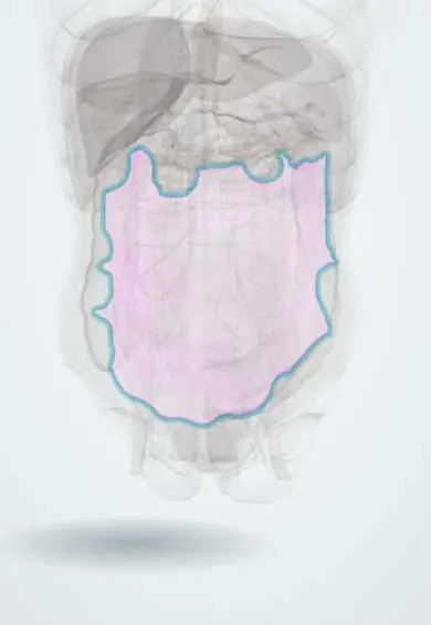
Structures involved
Tap one to see what it is and open it in 3D.
Microorganisms that cause it
Why it happens
- Patterns of acute inflammation: serous, fibrinous, purulent, abscess, ulcer
- Outcomes of acute inflammation: resolution, abscess, scar or chronic inflammation
- Serous membranes: pleura and peritoneum
What it is
A localised collection of pus within the peritoneal cavity, the retroperitoneum or an abdominal organ, walled off by inflamed bowel, omentum, mesentery and fibrin. Common sites are under the diaphragm (subphrenic), beside the colon (pericolic), between bowel loops, in the pelvis and in the iliopsoas muscle.
Who gets it
A frequent complication of complicated appendicitis, diverticulitis and abdominal surgery, especially after anastomotic leaks. In Colombia, late-presenting perforated appendicitis and abdominal trauma are important causes, and it is a common reason for re-operation or interventional radiology drainage.
Causes
- Perforated appendicitis or diverticulitis
- Postoperative leak or contamination (anastomotic leak, bowel injury)
- Generalised peritonitis that becomes localised
- Crohn disease (fistula and interloop or psoas abscess)
- Infected pancreatic necrosis, pelvic inflammatory disease (tubo-ovarian abscess)
- Penetrating or blunt abdominal trauma
- Psoas abscess: secondary to bowel, kidney or spine infection (including tuberculosis) or primary by blood-borne Staphylococcus aureus
Risk factors
- Delayed diagnosis of perforation
- Recent abdominal or pelvic surgery
- Immunosuppression, diabetes, malnutrition
- Corticosteroid or biologic therapy
- Injection drug use (primary psoas abscess)
- Older age and serious underlying illness
Pathophysiology
When bowel content or infection enters the peritoneal cavity, peritoneal defences (omentum, fibrin, phagocytes) try to contain it. If the bacterial load is not cleared, a capsule forms around a pocket of pus. Infection is usually polymicrobial from bowel flora: facultative gram-negative rods such as Escherichia coli and Klebsiella together with anaerobes, above all Bacteroides fragilis, which encourages abscess formation; enterococci and Candida appear in postoperative or immunocompromised patients. Because antibiotics penetrate the capsule poorly, the pus must usually be drained. Fluid flows by gravity and along peritoneal spaces, which explains collections in the subphrenic spaces, paracolic gutters and the pelvis.
Classification
- Intraperitoneal: subphrenic (right or left), subhepatic, paracolic, interloop, pelvic (pouch of Douglas)
- Retroperitoneal: pancreatic, perinephric, psoas (iliopsoas)
- Visceral: hepatic, splenic
- Primary psoas abscess (blood-borne) versus secondary (spread from a neighbouring infection)
Symptoms and signs
- Persistent or swinging fever with chills
- Abdominal pain and tenderness over the collection, sometimes a palpable mass
- Prolonged ileus, anorexia, nausea and malaise after surgery
- Subphrenic: shoulder-tip pain, hiccups, pleural effusion and basal lung changes
- Pelvic: diarrhoea, tenesmus, urinary frequency; tender mass on rectal or vaginal exam
- Psoas: flank or groin pain, limp, and pain on hip extension (psoas sign) with a flexed hip
Diagnosis
- Clinical
- Suspect it in any patient with fever, abdominal pain or failure to improve after perforation, peritonitis or abdominal surgery. Look for sepsis and organ dysfunction.
- Tests
- Full blood count (leukocytosis), C-reactive protein, lactate, kidney and liver function, blood cultures before antibiotics. Gram stain and culture of pus obtained at drainage to guide therapy.
- Imaging
- Contrast-enhanced CT of the abdomen and pelvis is the test of choice: it shows a rim-enhancing fluid collection, sometimes with gas, and guides drainage. Ultrasound is useful for subphrenic, hepatic and pelvic collections and in pregnancy or children; MRI is an alternative. Plain films may show air-fluid levels or loss of the psoas shadow.
Differential diagnosis
- Uncontained generalised peritonitis
- Postoperative haematoma or seroma, sterile fluid collection
- Pneumonia or empyema (for subphrenic collections)
- Pyelonephritis or perinephric abscess
- Pseudomembranous colitis or other cause of postoperative fever
- Tumour with central necrosis
Goal of treatment
Control the source by draining the pus and fixing the cause, treat the infection with suitable antibiotics, and support the patient through sepsis.
Treatment
- Acute
- Sepsis bundle when indicated: blood cultures, early broad-spectrum antibiotics, intravenous fluids and vasopressors as needed.
- Procedure
- Image-guided (CT or ultrasound) percutaneous drainage is first-line for most accessible single collections; very small pericolic or periappendiceal abscesses may settle with antibiotics alone.
- Surgical
- Laparoscopic or open surgery when drainage is not possible or fails, for multiple loculated collections, generalised peritonitis or to deal with the source (resection, repair of leak).
- Medical
- Antibiotics covering gram-negative bacilli and anaerobes (for example a beta-lactam with beta-lactamase inhibitor, a carbapenem, or a cephalosporin or fluoroquinolone plus metronidazole), narrowed by cultures, for a short course after adequate source control. Antituberculous therapy for tuberculous psoas abscess.
- Follow-up
- Nursing priorities: vital signs and temperature curve, drain output and character, drain patency and skin care, fluid balance, pain control, nutrition and early mobilisation.
Complications
- Sepsis and septic shock, multiple organ failure
- Rupture into the peritoneum causing generalised peritonitis
- Fistula to the bowel or skin
- Recurrent abscess or residual collection
- Pleural effusion or empyema (subphrenic)
- Bowel obstruction from adhesions
Prognosis
With prompt drainage and antibiotics most patients recover. Mortality rises with delayed source control, sepsis, older age and serious underlying disease rather than with the site of the abscess.
Prevention
- Early diagnosis and surgery of appendicitis and other perforations
- Appropriate surgical antibiotic prophylaxis and careful technique
- Thorough peritoneal toilet when the abdomen is contaminated
Test yourself
Think of the answer, then tap the question to see it.
What is the imaging test of choice for suspected intra-abdominal abscess?
Contrast-enhanced CT of the abdomen and pelvis, which also guides percutaneous drainage.
Which anaerobe is classically involved in intra-abdominal abscess formation?
Bacteroides fragilis, usually together with gram-negative rods such as Escherichia coli.
Why are antibiotics alone usually not enough for an abdominal abscess?
The walled-off pus is poorly penetrated by antibiotics, so source control by drainage is needed.
What clinical sign suggests a psoas abscess?
Pain on hip extension (psoas sign) with the hip held flexed, plus fever and flank or groin pain.
A patient has fever and shoulder-tip pain a week after a perforated duodenal ulcer repair. Where is the abscess likely?
In the subphrenic space, irritating the diaphragm (referred pain through the phrenic nerve).
Related conditions
- Peritonitis
- Acute appendicitis
- Diverticular disease and diverticulitis
- Crohn disease
- Acute pancreatitis
- Pelvic inflammatory disease
- Sepsis and septic shock
- Pyogenic liver abscess
- Amoebic liver abscess
- Vertebral osteomyelitis
Educational information, not medical advice. For any symptom, see a health professional.
Sources: Intra-Abdominal Abscesses - Gastroenterology - Merck Manual Professional Edition (Merck Manual Professional Edition, 2025); Diagnosis and Management of Complicated Intra-abdominal Infection in Adults and Children: Guidelines by the Surgical Infection Society and the Infectious Diseases Society of America (Clinical Infectious Diseases, 2010).
Spotted a mistake?We are always improving for the community. Tell us what is wrong and our team will review it.Report a mistake
Representative model, for educational purposes only.