
Lymphatic malformations (cystic hygroma)
A soft, spongy swelling in a baby's neck can be a pocket of lymph vessels that never joined up. It is benign, but in the neck it can squeeze the airway.
Quick facts
- Key signs
- Soft · Sudden enlargement · Clusters of small clear
- First line
- Under imaging guidance the cyst is drained
Red flags ↓
No doses: the clinician sets them by the current guideline.
What it is
A lymphatic malformation is a benign, slow-flow vascular malformation made of abnormal lymphatic channels and cysts filled with lymph that do not drain into the normal lymphatic system. It is present from before birth, grows with the child and does not involute on its own the way a haemangioma does. Most are in the head and neck (the old name cystic hygroma refers to the large-cyst type in the neck) or the axilla; they can also involve the mediastinum, the mesentery and the limbs.
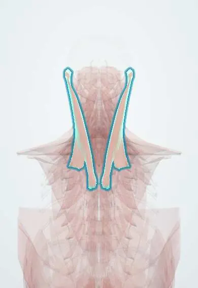
Who gets it
Uncommon: roughly 1 in several thousand births. About three quarters arise in the head and neck and about a fifth in the axilla; most are evident at birth or by age 2. Large neck cysts are now often seen on prenatal ultrasound. A posterior neck cystic hygroma found in the first trimester is a different, more ominous finding, strongly linked to chromosomal disorders (Turner syndrome, trisomies 21, 18 and 13), Noonan syndrome, heart defects and hydrops.
Causes
- Faulty development of the embryonic lymphatic sacs and channels, which fail to connect with the venous system
- Somatic (mosaic) activating mutations in the PIK3CA growth pathway in many lesions, especially microcystic and those in overgrowth syndromes
- First-trimester nuchal cystic hygroma: delayed lymphatic–jugular connection, often with aneuploidy
Risk factors
- Chromosomal disorders: Turner syndrome, trisomies 21, 18 and 13 (fetal nuchal hygroma)
- Noonan syndrome
- PIK3CA-related overgrowth syndromes
- No modifiable risk factors are known for isolated lesions
Pathophysiology
Abnormal lymphatic channels with no outflow → lymph accumulates in thin-walled cysts lined by lymphatic endothelium → the mass grows in proportion with the child and infiltrates between muscles, nerves and vessels rather than pushing them aside → infection (often after an upper respiratory infection) or bleeding into a cyst → rapid swelling, pain and redness → in the neck, floor of mouth or tongue, compression of the pharynx or trachea → stridor, feeding difficulty and airway obstruction. In the abdomen, leaking abnormal lymphatics fill the peritoneum with milky chyle (chylous ascites) → loss of protein, fat and lymphocytes.
Classification
- Macrocystic: one or more large cysts (roughly 1 cm or more); responds well to sclerotherapy
- Microcystic: many tiny cysts in a spongy mass, often with skin or mucosal vesicles; infiltrative, harder to treat
- Mixed macrocystic and microcystic
- Complex lymphatic anomalies (generalized lymphatic anomaly, Gorham–Stout disease with bone loss, central conducting lymphatic anomaly with chylous effusions)
- Part of combined vascular malformations or overgrowth syndromes (for example lymphatic–venous malformations)
Symptoms and signs
- Soft, painless, compressible, transilluminating swelling, most often in the posterior or anterior neck, face or axilla, present at birth or appearing in the first 2 years
- Sudden enlargement, firmness, pain and redness during a cold or after bleeding into a cyst
- Clusters of small clear or blood-tinged vesicles on the skin or tongue that leak fluid (microcystic)
- Enlarged tongue, drooling, difficulty feeding, snoring or sleep apnoea when the mouth or tongue are involved
- Stridor or breathing difficulty with neck and mediastinal lesions
- Chylous ascites: a distended abdomen with milky fluid in an infant, poor growth and oedema
Diagnosis
- Clinical
- A soft, compressible, non-pulsatile mass that transilluminates, does not change with crying or straining as a venous malformation does, and has been present since birth. Look at the mouth and tongue for vesicles and check the airway and feeding. In a fetus or newborn with a large neck mass, the first question is whether the airway is safe.
- Tests
- Aspirated fluid is clear or straw-coloured lymph (milky in chylous ascites, with high triglycerides and many lymphocytes). Biopsy is rarely needed; when done, the cyst lining stains for lymphatic endothelial markers. Genetic testing of fetal cells when a first-trimester nuchal hygroma is found.
- Imaging
- Ultrasound first: thin-walled, fluid-filled cysts with septa and no flow inside, sometimes with fluid–fluid levels after bleeding. MRI shows the full extent: cysts bright on T2-weighted images, with enhancement only of the walls and septa, and their relation to the airway, nerves and mediastinum. Prenatal ultrasound and fetal MRI plan delivery when the neck is involved.
Differential diagnosis
- Infantile haemangioma
- Venous malformation
- Branchial cleft cyst or thyroglossal duct cyst
- Cervical lymphadenitis or neck abscess
- Teratoma or other congenital neck tumour
- Lipoma or plexiform neurofibroma
Soft swellings in a baby's neck: how to tell them apart
| Lesion | When it shows and how it behaves | Feel and examination | Imaging clue |
|---|---|---|---|
| Lymphatic malformation | At birth or before 2 years; grows with the child, swells with infections; does not involute | Soft, compressible, transilluminates; skin vesicles if microcystic | Cysts with septa, no internal flow |
| Infantile haemangioma | Appears in the first weeks, grows fast for months, then slowly fades | Warm, firm-rubbery, bright red or bluish | Solid, high-flow mass with many vessels |
| Venous malformation | Present from birth, grows slowly; enlarges when dependent, crying or straining | Bluish, soft, empties on pressure | Slow-flow channels with phleboliths |
| Branchial cleft cyst | Usually noticed in older children or young adults, often after a cold | Single smooth cyst at the front edge of the sternocleidomastoid | One thick-walled cyst, no septa |
| Neck abscess | Days of fever and a growing painful lump | Hot, tender, red, the child is unwell | Collection with enhancing rim and inflamed tissue |
Goal of treatment
Keep the airway safe, treat infection and bleeding episodes, shrink or remove the malformation while preserving nerves and function, and control lymph leaks.
Treatment
- Acute
- Airway threat: secure the airway first; a fetus with a large neck lesion is delivered in a centre able to perform an ex utero intrapartum treatment (EXIT) procedure, securing the airway while the placenta still supports the baby.
- Medical
- Infection of the malformation: antibiotics and anti-inflammatory treatment; steroids are sometimes added for airway swelling. Treatment of the lesion itself is planned once inflammation settles.
- Procedure
- Sclerotherapy, first-line for macrocystic lesions: under imaging guidance the cyst is drained and a sclerosant (doxycycline, bleomycin, ethanol or OK-432) is injected so the walls scar and stick together; several sessions may be needed.
- Medical
- Microcystic, extensive or complex lesions: sirolimus, an mTOR inhibitor that blocks the PIK3CA pathway, can shrink them and reduce leakage; PIK3CA inhibitors are used in selected overgrowth syndromes in specialist centres.
- Surgical
- Surgical excision for well-localised lesions or those failing other treatment; complete removal is often impossible because the lesion wraps around nerves (facial, hypoglossal, vagus) and vessels, so partial resection that preserves function is accepted. Laser or radiofrequency ablation treats mucosal vesicles on the tongue.
- Lifestyle
- Chylous ascites or chylous effusions: a diet low in long-chain fat with medium-chain triglycerides (absorbed directly into the portal blood, bypassing the lymphatics) or parenteral nutrition; octreotide, drainage or surgery for persistent leaks.
Complications
- Airway obstruction and need for tracheostomy
- Recurrent infection (cellulitis of the malformation)
- Bleeding into cysts with sudden enlargement
- Feeding and speech difficulties, macroglossia, jaw overgrowth and dental malocclusion
- Cosmetic deformity
- Nerve injury after surgery; recurrence after incomplete excision
- Protein loss, malnutrition and immune deficiency from chylous ascites or chylothorax
Common traps
- A lymphatic malformation will fade like a haemangioma if you wait: it is a malformation, not a tumour, and grows with the child; it rarely disappears and swells during infections
- Surgery is always the first treatment: macrocystic lesions usually respond to sclerotherapy, and surgery risks damaging nerves the lesion wraps around
- A neck hygroma on a first-trimester scan is the same as a neck lymphatic malformation at birth: the fetal posterior nuchal hygroma is a marker of aneuploidy, heart defects and hydrops and needs genetic testing
- Every lymphatic malformation can be drained with a needle: microcystic lesions are a sponge of tiny cysts with nothing to aspirate; they are treated with sirolimus, laser or surgery
- Milky ascites in an infant means infection: chylous ascites from abnormal lymphatics is treated first with a diet of medium-chain fats, not antibiotics
Prognosis
Isolated macrocystic lesions respond well to sclerotherapy and have an excellent outlook. Microcystic and extensive cervicofacial lesions are a chronic condition with flare-ups and need long-term care. First-trimester nuchal hygroma with aneuploidy or hydrops carries a poor prognosis; isolated hygromas with normal chromosomes often resolve.
Prevention
- No primary prevention is known
- Detailed prenatal ultrasound, and fetal MRI for neck lesions, to plan delivery where the airway can be secured
- Genetic counselling and testing when a first-trimester nuchal hygroma is found
- Prompt treatment of upper respiratory infections and dental care in children with cervicofacial lesions
Test yourself
Think of the answer, then tap the question to see it.
A 2-year-old's soft neck swelling, present since birth, has doubled in size over 2 days during a cold. Why did it grow, and what do you check first?
Infection or bleeding into the cysts makes them swell quickly. Check the airway first (stridor, retractions, feeding), then treat the infection and plan definitive treatment later.
Why does a macrocystic lymphatic malformation respond to sclerotherapy while a microcystic one responds poorly?
Large cysts can be drained and injected so their walls scar together; a microcystic lesion is a sponge of tiny cysts the sclerosant cannot reach well, so sirolimus, laser or surgery are used.
How is a medium-chain triglyceride diet useful in chylous ascites?
Medium-chain fats are absorbed directly into portal venous blood, so they do not travel through the intestinal lymphatics; chyle flow and the leak fall while the infant still gets energy.
What does a posterior nuchal cystic hygroma at the 12-week scan prompt, and why?
Genetic testing and a detailed heart scan: it is strongly associated with Turner syndrome, trisomies, Noonan syndrome, heart defects and hydrops.
Connections
Educational information, not medical advice. For any symptom, see a health professional.
Sources: Lymphatic Malformations (StatPearls (NCBI Bookshelf), 2025); Nonimmune Hydrops Fetalis (StatPearls (NCBI Bookshelf), 2025); Ascites (StatPearls (NCBI Bookshelf), 2025); Lymphatic Diseases (MedlinePlus (U.S. National Library of Medicine), 2025).
Spotted a mistake?We are always improving for the community. Tell us what is wrong and our team will review it.Report a mistake
Representative model, for educational purposes only.