
Pancreatic cystic neoplasms and the incidental pancreatic cyst (IPMN, MCN, serous)
Scans often find small pancreatic cysts. Most are harmless, but some are early forms of cancer. The job is to tell which, without operating on too many.
What it is
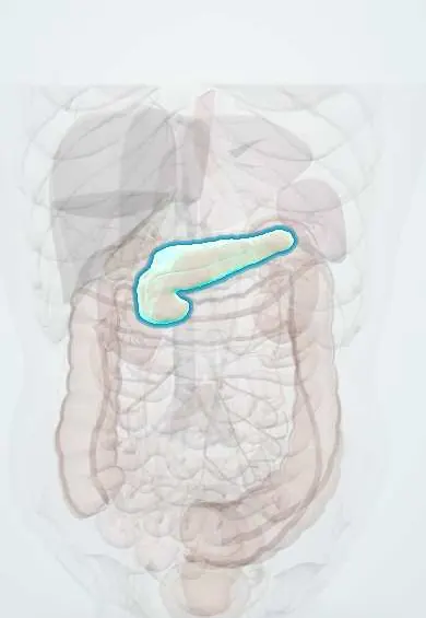
Fluid-filled lesions of the pancreas, most found by chance on CT or MRI done for another reason. The important group are cystic neoplasms: serous cystic neoplasms (serous cystadenoma, almost always benign), and the mucin-producing ones, intraductal papillary mucinous neoplasm (IPMN, which grows inside and communicates with the pancreatic ducts) and mucinous cystic neoplasm (MCN, which does not), both of which can progress through dysplasia to invasive cancer. Solid pseudopapillary neoplasm and cystic neuroendocrine tumours are rarer. Pseudocysts, which follow pancreatitis, are not neoplasms. Management uses imaging features grouped as 'worrying features' and 'high-risk stigmata' to decide between surveillance, endoscopic ultrasound and surgery.
Who gets it
Pancreatic cysts are found in a few per cent of adults who have abdominal CT, and in a higher share on MRI, rising steeply with age; most are small branch-duct IPMNs. Serous cystadenomas are typical of women in their sixties, MCNs of middle-aged women (almost exclusively), IPMNs of both sexes around the seventh decade, and solid pseudopapillary neoplasms of young women. As imaging use grows in Colombia, the incidental pancreatic cyst is a common reason for referral to surgery and gastroenterology.
Causes
- IPMN: papillary, mucin-secreting growth of duct epithelium; frequent GNAS and KRAS mutations
- MCN: mucin-secreting epithelium with ovarian-type stroma, probably from ectopic ovarian tissue during development, hence its strong female predominance
- Serous cystadenoma: glycogen-rich cuboidal epithelium; associated with von Hippel–Lindau disease
- Solid pseudopapillary neoplasm: CTNNB1 (beta-catenin) mutations
- Pseudocyst (not a neoplasm): walled-off collection of pancreatic juice after acute or chronic pancreatitis
Risk factors
- Older age (incidental cysts and IPMN)
- Female sex (MCN, serous cystadenoma, solid pseudopapillary neoplasm)
- Von Hippel–Lindau disease (serous cysts) and inherited pancreatic cancer syndromes
- Previous acute or chronic pancreatitis, alcohol and gallstones (pseudocyst)
- Family history of pancreatic cancer
Pathophysiology
Mucinous lesions follow a stepwise path: mutation in duct or cyst epithelium → mucin-secreting neoplasm → low-grade then high-grade dysplasia → invasive adenocarcinoma. In IPMN, mucin fills and dilates the ducts → main-duct involvement and mural nodules mark higher risk; in the head, a growing lesion can block the bile duct → jaundice; thick mucin can block the pancreatic duct → recurrent pancreatitis. MCN grows as a single thick-walled cyst in the body or tail, without duct connection. Serous cystadenoma grows slowly as many tiny cysts around a central scar and almost never becomes malignant, so it causes trouble only by size. A pseudocyst has no epithelial lining and follows pancreatitis.
Classification
- Serous cystic neoplasm (serous cystadenoma): microcystic 'honeycomb' with a central scar that may calcify; benign in nearly all
- Mucinous cystic neoplasm: single, thick-walled cyst in the body or tail of middle-aged women, no duct communication, ovarian-type stroma; malignant potential
- IPMN: main-duct (highest cancer risk), branch-duct (lowest, most common incidental cyst) or mixed type; communicates with the pancreatic duct
- Solid pseudopapillary neoplasm: mixed solid and cystic mass in young women; low-grade malignant
- Cystic neuroendocrine tumour and pseudocyst (non-neoplastic) as look-alikes
- Risk features (international consensus concepts): high-risk stigmata (obstructive jaundice with a head lesion, an enhancing solid component or mural nodule, markedly dilated main duct) and worrying features (larger cyst, thickened enhancing wall, moderately dilated main duct, small nodule, abrupt change in duct calibre, rising CA 19-9, rapid growth, pancreatitis)
Symptoms and signs
- Most have none and are found by chance
- Vague upper abdominal pain or back pain, early fullness
- Recurrent episodes of acute pancreatitis (mucin plugging the duct in IPMN)
- Jaundice, weight loss or new diabetes (warning signs of malignancy in a head lesion)
- Steatorrhoea from loss of exocrine function in extensive main-duct IPMN
- Palpable mass with very large serous or mucinous cysts or solid pseudopapillary neoplasm
Diagnosis
- Clinical
- Take the history that sorts the cyst: previous pancreatitis or alcohol (pseudocyst), sex and age (MCN in women, IPMN in older adults, solid pseudopapillary neoplasm in young women), symptoms such as jaundice, weight loss, new diabetes or steatorrhoea, family history and fitness for major surgery. A patient who would never be fit for or want pancreatic surgery may need no surveillance.
- Tests
- CA 19-9 as a supportive marker, liver tests if a head lesion or jaundice is present, and glucose or HbA1c for new diabetes. At endoscopic ultrasound, cyst fluid analysis: high CEA, string sign and low glucose suggest a mucinous cyst; very high amylase suggests connection with the duct (pseudocyst or IPMN); cytology looks for high-grade dysplasia or cancer; molecular markers (KRAS, GNAS) are used in some centres.
- Imaging
- MRI with MRCP is the best study to characterise a cyst and is the usual tool for surveillance: it shows duct communication, septations, nodules and the main duct calibre without radiation. Pancreas-protocol contrast CT is an alternative and shows calcification (the central scar of a serous cystadenoma, the peripheral eggshell rim of some MCNs). Endoscopic ultrasound gives the most detailed view of walls and nodules, allows contrast enhancement of a nodule and guides fine-needle aspiration; it is used for cysts with worrying features or uncertain type.
Differential diagnosis
- Pancreatic pseudocyst or walled-off necrosis after pancreatitis
- Pancreatic ductal adenocarcinoma with cystic degeneration
- Cystic pancreatic neuroendocrine tumour
- Simple (true) epithelial cyst, lymphoepithelial cyst
- Duodenal diverticulum or choledochal cyst seen next to the pancreas
- Cysts in polycystic kidney or von Hippel–Lindau disease
Pancreatic cyst: which one is it?
| Lesion | Typical patient and site | Imaging clue | Fluid and risk |
|---|---|---|---|
| Pseudocyst | History of pancreatitis or alcohol; anywhere | Single cyst, no septa, often with signs of pancreatitis | Very high amylase, low CEA; not a neoplasm |
| Serous cystadenoma | Woman in her sixties; often the head | Honeycomb of tiny cysts with a central scar or calcification | Low CEA; benign in nearly all |
| Mucinous cystic neoplasm | Woman in middle age; body or tail | Single thick-walled cyst, no duct connection, sometimes rim calcification | High CEA; premalignant, usually resected |
| IPMN | Man or woman over 60; often the head | Cyst or dilated duct that connects with the main duct; nodules are a warning | High CEA and amylase; risk depends on duct involvement and features |
| Solid pseudopapillary neoplasm | Young woman; body or tail | Mixed solid and cystic mass, sometimes bleeding inside | Low-grade malignant; resected |
| Cystic neuroendocrine tumour | Adult, sometimes with MEN1 | Cyst with a brightly enhancing rim in the arterial phase | Low CEA; malignant potential |
Goal of treatment
Find the few cysts that are or will become cancer and remove them at the right time, while sparing the many harmless cysts from surgery, and stopping surveillance in patients who would not benefit from an operation.
Treatment
- Follow-up
- Small branch-duct IPMNs and indeterminate cysts without worrying features: surveillance with MRI and MRCP, with intervals set by size and stability and lengthened when the cyst stays stable; surveillance stops when the patient is no longer fit for or willing to have surgery.
- Procedure
- Cysts with worrying features or uncertain type: endoscopic ultrasound with fine-needle aspiration for fluid analysis and cytology, and contrast assessment of any nodule; the result moves the cyst either to surgery or back to surveillance.
- Surgical
- High-risk stigmata, main-duct IPMN, mucinous cystic neoplasm, solid pseudopapillary neoplasm and symptomatic cysts in fit patients: oncological pancreatic resection by an experienced team: pancreaticoduodenectomy (Whipple) for the head, distal pancreatectomy, often with splenectomy, for the body and tail; occasionally total pancreatectomy for diffuse main-duct disease.
- Conservative
- Serous cystadenoma with a typical appearance: no surveillance or limited follow-up; resection only if it causes symptoms or compression, or if the diagnosis is in doubt.
- Follow-up
- After resection of an IPMN: continued imaging of the remaining pancreas, because new or recurrent lesions can develop elsewhere in the gland. After pancreatectomy: management of exocrine insufficiency with pancreatic enzymes and of diabetes.
Complications
- Progression to invasive pancreatic adenocarcinoma (mucinous lesions)
- Recurrent acute pancreatitis
- Obstructive jaundice
- Exocrine insufficiency and diabetes, from the disease or after resection
- Pancreatic fistula, delayed gastric emptying and bleeding after pancreatic surgery
- Harms of overtreatment: major surgery for cysts that would never have become cancer
- Anxiety and costs of unnecessary or prolonged surveillance
Common traps
- Every pancreatic cyst is a pseudocyst: without a history of pancreatitis, an incidental cyst is most often a neoplasm, usually a branch-duct IPMN, and needs proper characterisation
- Every cystic neoplasm should be removed: most small branch-duct IPMNs never become cancer, serous cystadenomas are benign, and pancreatic surgery has real morbidity; risk features, not the cyst itself, drive the decision
- A cyst with a solid enhancing nodule can be watched like the others: an enhancing mural nodule is a high-risk stigma and points to surgery in a fit patient
- A low CEA in the fluid rules out a mucinous cyst: CEA is specific but not sensitive; low glucose, cytology, imaging and molecular markers add to the picture
- Surveillance never stops: it stops when the patient is no longer a candidate for surgery, because there is nothing left to decide
Prognosis
Most incidental cysts, especially small branch-duct IPMNs without worrying features, stay stable for years and rarely become cancer. Serous cystadenomas are benign. Resected MCNs and IPMNs without invasion are usually cured, although IPMN patients need lifelong follow-up of the remaining gland. Once invasive cancer develops, the outlook approaches that of pancreatic adenocarcinoma, although cancers from IPMN found during surveillance tend to be caught earlier.
Prevention
- No proven way to prevent cyst formation; avoiding alcohol and treating gallstones lowers the risk of pancreatitis and pseudocysts
- Structured surveillance of mucinous cysts so that worrying changes are found before cancer spreads
- Refer every incidental pancreatic cyst to a pancreatic team or protocol instead of ad hoc scans
- Screening programmes for people at high inherited risk of pancreatic cancer
Test yourself
Think of the answer, then tap the question to see it.
A 45-year-old woman has a single thick-walled 4 cm cyst in the tail of the pancreas, not connected to the duct, with high CEA in the fluid. What is it, and what is advised?
A mucinous cystic neoplasm: middle-aged woman, body or tail, no duct communication, mucinous fluid. It is premalignant, so distal pancreatectomy is usually advised in a fit patient.
Why is main-duct IPMN treated more aggressively than branch-duct IPMN?
Main-duct involvement carries a much higher risk of high-grade dysplasia or invasive cancer; most small branch-duct IPMNs stay low risk and can be watched.
Which concepts make up the high-risk stigmata of a mucinous pancreatic cyst?
Obstructive jaundice with a lesion in the head of the pancreas, an enhancing solid component or mural nodule, and a markedly dilated main pancreatic duct.
A 66-year-old woman has a microcystic honeycomb lesion with a calcified central scar. Why can she usually avoid surgery?
It is a serous cystadenoma, which is benign in nearly all cases; it is removed only if it causes symptoms or the diagnosis is uncertain.
An 88-year-old with heart failure has a 15 mm branch-duct IPMN. Why might no surveillance be the best plan?
Surveillance only helps if a worrying change would lead to surgery; a patient who could not safely have a pancreatectomy gains nothing and bears the cost and anxiety.
Why it happens
- Cystic pancreatic neoplasms
- Non-neoplastic pancreatic cysts: congenital cysts and pseudocysts
- Precursors of pancreatic cancer: PanIN, IPMN, MCN
Connections
Educational information, not medical advice. For any symptom, see a health professional.
Sources: Intraductal Papillary Mucinous Neoplasm of the Pancreas (StatPearls (NCBI Bookshelf), 2026); Mucinous Cystic Pancreatic Neoplasms (StatPearls (NCBI Bookshelf), 2026); Serous Cystic Neoplasms of the Pancreas (StatPearls (NCBI Bookshelf), 2026); Pancreatic Pseudocyst (StatPearls (NCBI Bookshelf), 2026).
Spotted a mistake?We are always improving for the community. Tell us what is wrong and our team will review it.Report a mistake
Representative model, for educational purposes only.