
Duodenal and jejunal diverticula
Small pouches can bulge out of the small bowel wall. Most never cause harm, but a few bleed, burst, block the bile duct or breed bacteria.
What it is
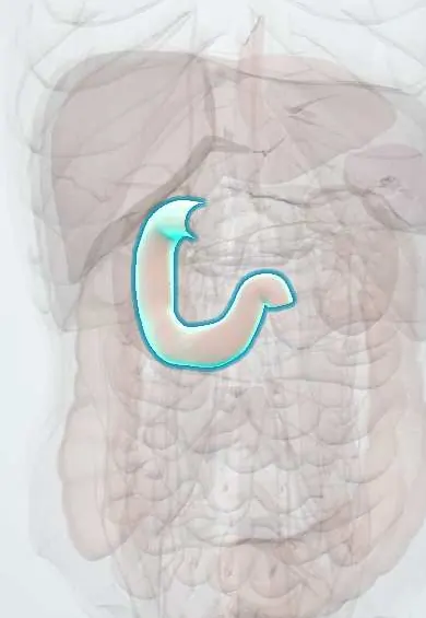
Outpouchings of the wall of the small bowel. Duodenal diverticula are common, usually single and lie on the inner (medial) wall of the second part of the duodenum, most within a few centimetres of the papilla (periampullary). Jejunal and ileal diverticula are rarer, usually multiple and sit on the mesenteric border. Both are acquired false (pulsion) diverticula made of mucosa and submucosa without muscle, unlike the congenital, true Meckel diverticulum.
Who gets it
Duodenal diverticula are found in a sizeable minority of people having ERCP and in many older adults on CT; they become commoner with age and are rare before 40. Jejunoileal diverticulosis is much rarer and seen mostly in older adults. The great majority never cause symptoms. In Colombia, where gallstone disease and ERCP are common, a periampullary diverticulum matters mainly because it makes cannulation of the papilla harder and favours recurrent bile-duct stones.
Causes
- Acquired pulsion: pressure inside the bowel pushes mucosa through weak points where vessels or ducts pierce the muscle layer
- Disorders of small bowel motility (visceral myopathy or neuropathy, systemic sclerosis) in jejunal diverticulosis
- Age-related weakening of the bowel wall
- Rarely, a congenital intraluminal (windsock) duodenal diverticulum
Risk factors
- Older age
- Systemic sclerosis and other small bowel motility disorders
- Previous duodenal ulcer scarring (pseudodiverticula of the duodenal bulb)
Pathophysiology
Peristaltic pressure pushes mucosa and submucosa through a weak point where a vessel or duct enters the wall → a thin pouch without muscle forms. In the duodenum, a pouch beside the papilla distorts or compresses the end of the bile and pancreatic ducts and holds food and bile → stasis → brown pigment stones in the bile duct, cholangitis and pancreatitis, or obstructive jaundice without stones (Lemmel syndrome). In the jejunum, pouches without muscle empty poorly → stagnant contents → bacterial overgrowth → bacteria deconjugate bile acids and consume vitamin B12 → fatty diarrhoea, weight loss and B12 deficiency. At either site, inflammation of the thin wall → diverticulitis and perforation, often into the retroperitoneum; erosion of a vessel at the neck → bleeding.
Classification
- By site: duodenal (mostly periampullary, second part), jejunoileal (multiple, mesenteric border), and the separate congenital Meckel diverticulum (true, ileum)
- Periampullary: papilla inside the diverticulum, at its rim, or close beside it
- Extraluminal (acquired, the usual type) vs intraluminal (congenital windsock, rare)
- Uncomplicated (incidental) vs complicated: bleeding, diverticulitis or perforation, biliary or pancreatic disease, bacterial overgrowth, obstruction
Symptoms and signs
- Usually none: found on endoscopy, ERCP or CT done for another reason
- Vague upper abdominal pain, bloating or fullness after meals
- Jaundice, fever and right upper quadrant pain (cholangitis) or pancreatitis from a periampullary pouch
- Diarrhoea, fatty stools, weight loss and anaemia from bacterial overgrowth in jejunal diverticulosis
- Vomiting of blood, black stools or maroon stools from a bleeding diverticulum
- Sudden abdominal or back pain with fever when a diverticulum perforates
Diagnosis
- Clinical
- Most are incidental and need nothing. Think of them when bile-duct stones, cholangitis or pancreatitis recur after cholecystectomy, when an older adult has unexplained bacterial overgrowth or B12 deficiency, or when the source of small bowel bleeding or a retroperitoneal perforation is unclear.
- Tests
- Liver tests and lipase in biliary or pancreatic presentations, with blood cultures in cholangitis; full blood count, vitamin B12, folate (often high in overgrowth) and albumin; glucose or lactulose hydrogen breath test, or culture of jejunal aspirate, for overgrowth. Endoscopy with a side-viewing duodenoscope, capsule endoscopy or device-assisted enteroscopy show the pouches and any bleeding point.
- Imaging
- CT with oral and intravenous contrast: a gas- or fluid-filled sac beside the second part of the duodenum, against the pancreatic head, which can mimic a pancreatic cyst or abscess; oral contrast or gas inside it shows that it opens into the duodenum. Perforation shows retroperitoneal gas and fluid. Upper GI contrast or CT or MR enterography shows multiple jejunal pouches. MRCP shows a periampullary diverticulum and its relation to the bile duct. CT angiography or a tagged red cell scan localises bleeding.
Differential diagnosis
- Pancreatic cyst or pseudocyst
- Duodenal ulcer
- Bile-duct stones from the gallbladder or retained after cholecystectomy
- Meckel diverticulum
- Small bowel tumour
- Other causes of bacterial overgrowth (blind loop after surgery, motility disorders)
Pouches of the gut: which one?
| Type | Where | Wall | Typical trouble |
|---|---|---|---|
| Duodenal (periampullary) | Inner wall of the second part, beside the papilla; usually single | False: mucosa and submucosa, no muscle; acquired | Bile-duct stones, cholangitis, pancreatitis, difficult ERCP; rarely bleeding or perforation |
| Jejunoileal diverticulosis | Mesenteric border of the jejunum; usually multiple | False; acquired, older adults | Bacterial overgrowth, B12 deficiency, bleeding, perforation |
| Meckel diverticulum | Antimesenteric border of the distal ileum | True: all layers; congenital | Painless bleeding in children from gastric mucosa inside it, intussusception, obstruction |
| Colonic diverticula | Mostly sigmoid colon | False; acquired | Diverticulitis and lower GI bleeding |
Goal of treatment
Leave silent diverticula alone; treat each complication, preferring drugs and endoscopy, and keep surgery for free perforation, uncontrolled bleeding or repeated trouble.
Treatment
- Conservative
- Incidental diverticula need no treatment and no follow-up; the report should mention a periampullary diverticulum so a future endoscopist can plan.
- Medical
- Bacterial overgrowth: antibiotic courses, sometimes rotated, with vitamin B12 replacement and nutritional support. Cholangitis and pancreatitis are treated as usual, with antibiotics for cholangitis.
- Procedure
- ERCP to clear bile-duct stones or drain cholangitis, adapting sphincterotomy or balloon dilatation to where the papilla lies; endoscopic clips, injection or heat for a bleeding diverticulum; angiographic embolisation if endoscopy fails.
- Acute
- Perforation: resuscitation, nil by mouth, broad-spectrum antibiotics and a nasogastric tube; selected stable patients with a contained retroperitoneal leak can be managed this way with image-guided drainage and close surgical review.
- Surgical
- Surgery for free perforation, uncontrolled bleeding, obstruction or recurrent disease: excision of the diverticulum with repair of the duodenum, or resection of the affected segment of jejunum. Operations beside the papilla are difficult and risk injury to the bile and pancreatic ducts.
Complications
- Bleeding
- Diverticulitis and perforation, with retroperitoneal abscess
- Bile-duct stones, cholangitis, pancreatitis and obstructive jaundice (Lemmel syndrome)
- Bacterial overgrowth with malabsorption and vitamin B12 deficiency
- Stones formed inside a pouch (enteroliths) causing obstruction
- Perforation or failed cannulation during ERCP
Common traps
- A duodenal diverticulum seen on CT needs surgery: most are incidental and harmless; act only on a complication
- Duodenal diverticula are like Meckel diverticulum: Meckel is a true congenital pouch on the antimesenteric ileum; duodenal and jejunal ones are acquired false pouches on the medial or mesenteric side
- Recurrent bile-duct stones after cholecystectomy always mean a stone was left behind: a periampullary diverticulum causes stasis and new brown pigment stones, and can block the bile duct without any stone (Lemmel syndrome)
- A fluid-filled sac next to the pancreatic head is a pancreatic cyst: a duodenal diverticulum can look the same on CT; oral contrast or gas inside the sac shows it opens into the duodenum
- Low B12 with high folate in an older adult simply means a poor diet: that pattern points to bacterial overgrowth, and jejunal diverticulosis is a classic hidden cause
Prognosis
Excellent for incidental diverticula. Complications are uncommon, but perforation and heavy bleeding carry real mortality, mainly because the diagnosis is delayed in older patients with few signs.
Prevention
- No measure prevents them; report a periampullary diverticulum on ERCP, MRCP or CT so later procedures can be planned
- Consider a diverticulum when biliary disease recurs after cholecystectomy
- Treat bacterial overgrowth and replace vitamin B12 early in jejunal diverticulosis
Test yourself
Think of the answer, then tap the question to see it.
A 75-year-old has diarrhoea, weight loss, low vitamin B12 and high folate. CT shows many pouches along the jejunum. What links these findings?
Jejunal diverticulosis with bacterial overgrowth: stagnant pouches let bacteria multiply; they consume B12, make folate and deconjugate bile acids, causing fat malabsorption.
Why can a periampullary diverticulum cause bile-duct stones and cholangitis even after cholecystectomy?
The pouch beside the papilla compresses the end of the bile duct and holds food and bacteria, causing stasis, infection and new brown pigment stones inside the duct.
What is Lemmel syndrome?
Obstructive jaundice caused by a periampullary duodenal diverticulum pressing on the distal bile duct, with no stone or tumour.
Why is a perforated duodenal diverticulum easy to miss?
Most lie against the retroperitoneum, so the leak goes behind the peritoneum: there is little free gas under the diaphragm and few peritoneal signs, and the diagnosis needs CT.
Connections
Educational information, not medical advice. For any symptom, see a health professional.
Sources: Duodenal Perforation (StatPearls (NCBI Bookshelf), 2026); Small Intestinal Bacterial Overgrowth (StatPearls (NCBI Bookshelf), 2023); Cholangitis (StatPearls (NCBI Bookshelf), 2023).
Spotted a mistake?We are always improving for the community. Tell us what is wrong and our team will review it.Report a mistake
Representative model, for educational purposes only.