Conditions · Surgery and trauma
Traumatic diaphragmatic injury !
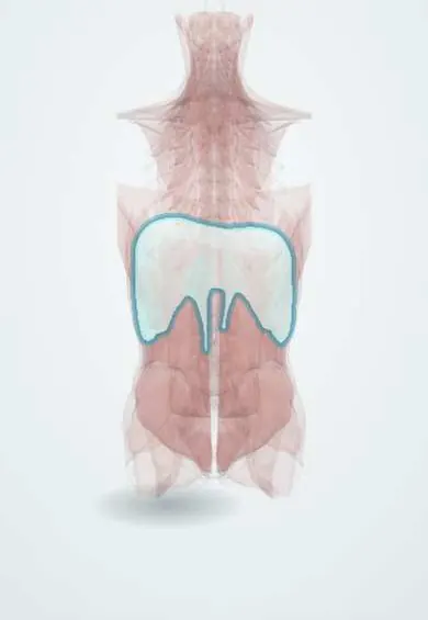
A traumatic diaphragm injury is a tear in the breathing muscle after blunt or penetrating trauma. Abdominal organs can slide into the chest, even years later.
Updated 2026-11-01
Structures involved
Tap one to see what it is and open it in 3D.
- Diaphragm
- Stomach
- Phrenic nerves
- Pleura
- Ribs (back part)
- Spleen
- Liver
- Transverse colon
- Greater omentum
- Esophagus
Why it happens
What it is
A tear or laceration of the diaphragm caused by blunt or penetrating trauma. It is a marker of severe trauma and is easily missed because it is often silent at first. Through the defect, abdominal organs (most often the stomach, colon, omentum and spleen) can herniate into the chest immediately or months to years later, causing breathing problems or bowel obstruction and strangulation.
Who gets it
Diaphragm injuries occur in a small proportion of trauma patients, more often after penetrating than blunt trauma. Left-sided injuries predominate because the liver shields the right hemidiaphragm. In Colombia and Latin America, stab and gunshot wounds of the lower chest and upper abdomen and motorcycle crashes are typical mechanisms. A substantial share of injuries are diagnosed late.
Causes
- Penetrating stab or gunshot wounds of the thoracoabdominal region (below the nipple line in front, below the tip of the scapula behind)
- High-speed motor vehicle and motorcycle crashes, especially lateral impact
- Falls from height
- Crush injuries and direct abdominal blows
Risk factors
- Penetrating wounds of the lower chest or upper abdomen, particularly on the left
- High-energy blunt trauma with pelvic or rib fractures
- Associated solid-organ injury (spleen, liver)
- Delayed presentation after previous trauma
Pathophysiology
In blunt trauma, a sudden rise in intra-abdominal pressure bursts the diaphragm, usually producing a long radial tear in the posterolateral left hemidiaphragm. Penetrating wounds create small holes that may not herniate at first. The pressure gradient between the abdomen (positive) and the chest (negative during inspiration) progressively pulls abdominal viscera through the defect. Herniated organs compress the lung and shift the mediastinum, and the bowel or stomach may become incarcerated, obstructed or strangulated. A right-sided injury implies greater force and frequent liver or major vascular injury.
Classification
- By mechanism: blunt (large tears) versus penetrating (small lacerations)
- By side: left (most common), right, bilateral (rare)
- By phase (Grimes): acute, latent (intermittent vague symptoms) and obstructive (incarceration or strangulation)
- AAST organ injury scale grades I–V based on the size of the tear and tissue loss
Symptoms and signs
- Chest or upper abdominal pain, sometimes referred to the shoulder
- Shortness of breath
- Bowel sounds heard in the chest and reduced breath sounds on the affected side
- Often no specific signs, with symptoms masked by other injuries
- In late presentation: nausea, vomiting, inability to pass stool or gas, and post-meal chest discomfort
Diagnosis
- Clinical
- ATLS primary survey. Suspect it in any thoracoabdominal penetrating wound and in high-energy blunt trauma. Physical examination is unreliable; a high index of suspicion is key, including in patients with remote trauma who present with obstruction or breathing symptoms.
- Tests
- No specific blood tests; trauma laboratory studies and blood gases as needed.
- Imaging
- Chest X-ray is the first test and may show an elevated or obscured hemidiaphragm, viscera or a nasogastric tube above it, but it is often normal. Contrast CT with multiplanar reconstruction is the main non-invasive test (collar sign, dependent viscera sign, discontinuity of the diaphragm). FAST helps detect associated bleeding. Diagnostic laparoscopy is the most accurate method in stable patients with left thoracoabdominal penetrating wounds.
Differential diagnosis
- Eventration or paralysis of the diaphragm
- Congenital (Bochdalek or Morgagni) diaphragmatic hernia
- Hiatal hernia
- Haemothorax, pneumothorax or pulmonary contusion
- Atelectasis and subpulmonary effusion
Goal of treatment
Find the defect, return the herniated organs to the abdomen and repair the diaphragm before strangulation or respiratory compromise occurs, while treating associated injuries.
Treatment
- Acute
- ATLS resuscitation, airway and oxygen support, chest drain for haemothorax or pneumothorax placed with care (herniated bowel can be injured), and nasogastric decompression of a herniated stomach.
- Surgical
- All diagnosed traumatic diaphragm injuries are repaired, because they do not heal and tend to enlarge. Acute injuries are usually repaired through the abdomen (laparotomy, or laparoscopy in stable patients), with non-absorbable sutures; mesh for large defects. Thoracoscopy is useful for some right-sided and chronic injuries.
- Procedure
- Diagnostic laparoscopy in haemodynamically stable patients with a left thoracoabdominal stab wound and no peritonitis, which can also be used for repair.
- Follow-up
- Nursing priorities: respiratory status and oxygen saturation, chest drain care, nasogastric tube function, pain control for deep breathing, early mobilisation and incentive spirometry; watch for signs of bowel obstruction.
Complications
- Delayed herniation of abdominal viscera into the chest
- Bowel or gastric incarceration, strangulation and perforation
- Tension gastrothorax and respiratory failure
- Pneumonia, empyema and atelectasis
- Recurrence after repair
Prognosis
Outcome largely depends on associated injuries; mortality in these patients is substantial because diaphragm injury marks high-energy trauma. Isolated repaired injuries do well. Delayed diagnosis with strangulated viscera carries a high risk of death.
Prevention
- Road safety: seatbelts and motorcycle safety measures
- Violence-prevention programmes
- Systematic evaluation of all left thoracoabdominal penetrating wounds
Test yourself
Think of the answer, then tap the question to see it.
Which side of the diaphragm is more often injured in blunt trauma, and why?
The left, because the liver protects the right hemidiaphragm.
Which herniated organ is most often found in the chest after left-sided diaphragm rupture?
The stomach.
What chest X-ray finding strongly suggests diaphragmatic rupture?
A nasogastric tube or the stomach seen above the left hemidiaphragm.
What is the preferred diagnostic method in a stable patient with a left thoracoabdominal stab wound?
Diagnostic laparoscopy.
Why must even small traumatic diaphragm tears be repaired?
They do not heal on their own and can enlarge, leading to delayed herniation and strangulation.
Related conditions
- Blunt abdominal trauma
- Penetrating trauma (stab and gunshot wounds)
- Major trauma (polytrauma): ATLS primary survey
- Splenic injury
- Liver injury
- Haemothorax
- Pneumothorax
- Rib fractures
- Congenital diaphragmatic hernia
- Hiatal hernia
- Small bowel obstruction
Educational information, not medical advice. For any symptom, see a health professional.
Sources: Traumatic diaphragmatic injury: a narrative review (Current Challenges in Thoracic Surgery, 2025); A review on delayed presentation of diaphragmatic rupture (World Journal of Emergency Surgery, 2009).
Spotted a mistake?We are always improving for the community. Tell us what is wrong and our team will review it.Report a mistake
Representative model, for educational purposes only.