
Cystic liver lesions: simple cyst, polycystic liver, mucinous cystic neoplasm, hydatid cyst and abscess
Fluid-filled spots in the liver are common and usually harmless, but a few are parasites, infections or growths that can become cancer.
What it is
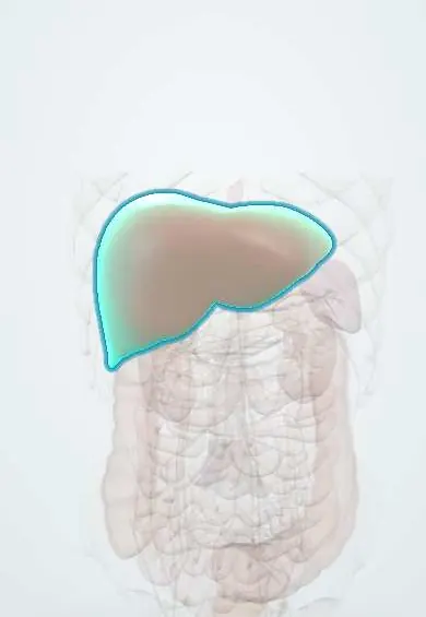
Any fluid-filled lesion of the liver seen on imaging. Most are simple cysts: thin-walled, filled with clear fluid, lined by bile-duct-type cells, not connected to the bile ducts and harmless. The others matter because each is treated differently: polycystic liver disease (many cysts, often with polycystic kidneys), mucinous cystic neoplasm (a premalignant tumour with internal septa, almost only in women), hydatid cyst (Echinococcus), abscess (pyogenic or amoebic), and cystic change in tumours or metastases. Two biliary problems are covered briefly here: haemobilia (bleeding into the bile ducts) and recurrent pyogenic cholangitis (repeated infection around pigment stones in the ducts inside the liver).
Who gets it
Simple cysts appear in roughly 2 to 18 percent of adults depending on the imaging method, more in women and with age. Polycystic liver disease is mostly part of autosomal dominant polycystic kidney disease, in which most adults have liver cysts by middle age, larger in women who have had pregnancies or taken oestrogens. Mucinous cystic neoplasms are rare. In Colombia, cystic echinococcosis is sporadic, while the polycystic form caused by Echinococcus vogeli occurs in rural and Amazon areas; amoebic abscess follows poor sanitation; recurrent pyogenic cholangitis is mostly an East Asian disease but appears in migrants and with liver flukes.
Causes
- Simple cyst: congenital, from tiny bile-duct remnants that lost contact with the biliary tree
- Polycystic liver disease: autosomal dominant polycystic kidney disease (PKD1, PKD2) or isolated autosomal dominant polycystic liver disease (PRKCSH, SEC63 and other genes)
- Mucinous cystic neoplasm: a tumour with mucin-secreting lining and ovarian-type stroma, almost only in women
- Hydatid cyst: larvae of Echinococcus granulosus from eggs in dog faeces; in the Americas also Echinococcus vogeli (polycystic echinococcosis)
- Abscess: bacteria arriving through the bile ducts or the portal vein (pyogenic) or Entamoeba histolytica (amoebic)
- Others: cystic metastases, biloma or haematoma after trauma or surgery, Caroli disease and choledochal cysts
Risk factors
- Female sex and older age (simple cysts)
- Family history of polycystic kidney or liver disease
- Pregnancies and oestrogen therapy (growth of polycystic liver)
- Close contact with dogs fed raw offal from livestock (hydatid)
- Biliary disease, diabetes and older age (pyogenic abscess); poor sanitation (amoebic abscess)
Pathophysiology
Simple cyst and polycystic liver: a fault in bile-duct development leaves pockets of bile-duct lining cut off from the duct system → the lining keeps secreting fluid → the cyst slowly fills and grows; in polycystic disease hundreds do this, and oestrogens speed it up → a huge liver presses on the stomach, diaphragm and veins (early fullness, breathlessness, rarely blocked venous outflow). Mucinous cystic neoplasm: a true tumour of mucin-secreting cells → septa and nodules → may turn into invasive carcinoma. Hydatid: swallowed eggs hatch → the larva reaches the liver through the portal vein → grows slowly inside a fibrous capsule made by the host, forming daughter cysts → rupture into the bile ducts (cholangitis, jaundice) or the peritoneum (anaphylaxis and spread). Abscess: infection reaches the liver through bile ducts, portal blood or the hepatic artery → tissue liquefies → fever, pain, sepsis. Haemobilia: an injured hepatic artery branch (after trauma, biopsy or drainage) forms a pseudoaneurysm that bleeds into a duct → pain, jaundice and gastrointestinal bleeding.
Classification
- Simple cyst vs complex cyst (septa, thick wall, nodules, calcification, debris)
- Polycystic liver, Gigot types: I few large cysts; II many medium cysts with normal liver between; III diffuse small and medium cysts with little normal liver
- Hydatid cyst, WHO ultrasound stages: CE1–CE2 active, CE3 transitional, CE4–CE5 inactive
- Neoplastic: mucinous cystic neoplasm (with low- or high-grade dysplasia, or invasive carcinoma) and cystic metastasis
- Infective: pyogenic abscess, amoebic abscess, infected cyst
Symptoms and signs
- Usually none: found on ultrasound or CT done for another reason
- Right upper quadrant fullness, dull pain, early satiety or a palpable liver when cysts are large or many
- Sudden severe pain from bleeding into a cyst or rupture
- Fever, rigors and a tender liver (abscess or infected cyst)
- Jaundice and cholangitis when a hydatid cyst opens into the bile ducts or a cyst compresses them
- Hives, wheeze or anaphylaxis when a hydatid cyst leaks
Diagnosis
- Clinical
- Most cysts are incidental: the task is to classify, not to treat. Ask about fever, travel and contact with dogs or livestock, family history of kidney cysts, recent liver procedures or trauma, and, in women, pregnancies and hormone use.
- Tests
- Liver tests (usually normal with simple cysts); full blood count and inflammatory markers if febrile, with blood cultures in abscess; serology for Echinococcus and Entamoeba histolytica; kidney function and family screening in polycystic disease. Aspiration of a possibly hydatid or mucinous cyst for diagnosis is avoided.
- Imaging
- Ultrasound first: a round, black (anechoic) lesion with an invisible wall, posterior acoustic enhancement and no septa or flow is a simple cyst and needs no follow-up. Contrast CT or MRI for anything complex: enhancing septa or wall nodules suggest a mucinous neoplasm; daughter cysts, a detached floating membrane (water-lily sign) or a calcified wall suggest hydatid; a thick enhancing rim with surrounding oedema, sometimes gas, suggests abscess. MRCP shows any link with the bile ducts and the stones and strictures of recurrent pyogenic cholangitis. In haemobilia, CT angiography and then angiography find the bleeding vessel or pseudoaneurysm.
Differential diagnosis
- Cystic metastasis (ovary, colon, neuroendocrine tumours)
- Biloma or haematoma after trauma or surgery
- Caroli disease and choledochal cyst
- Haemangioma or other solid lesion that looks cystic on ultrasound
- Necrotic hepatocellular carcinoma or cholangiocarcinoma
A cyst in the liver: which one?
| Lesion | Who | Imaging clue | What to do |
|---|---|---|---|
| Simple cyst | Anyone; more women, older adults | Invisible wall, no septa or nodules, posterior enhancement | Nothing, unless large and causing symptoms |
| Polycystic liver | Family history, polycystic kidneys | Dozens of cysts of many sizes, large liver | Treat symptoms: stop oestrogens, somatostatin analogues, fenestration, resection or transplant |
| Mucinous cystic neoplasm | Middle-aged women | Thick wall, enhancing septa, wall nodules | Complete surgical resection |
| Hydatid cyst | Contact with dogs and livestock | Daughter cysts, floating membrane, calcified wall | By stage: albendazole, PAIR or surgery |
| Pyogenic abscess | Biliary disease, diabetes, older adults | Thick enhancing rim, clustered cavities, sometimes gas | Antibiotics plus drainage |
| Amoebic abscess | Young men, poor sanitation | Single round lesion, usually right lobe under the diaphragm | Metronidazole then a luminal agent; drainage rarely |
Goal of treatment
Leave simple cysts alone, relieve the symptoms of large or many cysts, remove anything that may be a neoplasm, and treat parasites and infections with the specific approach each needs.
Treatment
- Conservative
- Asymptomatic simple cysts: no treatment and no follow-up imaging once confidently characterised.
- Surgical
- Large symptomatic simple cyst: laparoscopic deroofing (fenestration), which removes the cyst roof so the fluid drains into the peritoneum; aspiration alone almost always refills. Mucinous cystic neoplasm: complete resection, never fenestration, because the lining left behind can recur and become malignant.
- Procedure
- Image-guided aspiration with alcohol sclerotherapy for selected symptomatic cysts when surgery is unsuitable; percutaneous drainage of large pyogenic abscesses; PAIR (puncture, aspiration, injection of a scolicidal agent, re-aspiration) for suitable hydatid cyst stages under albendazole cover.
- Medical
- Polycystic liver: stop oestrogens; somatostatin analogues can slow growth. Hydatid: albendazole around any procedure, and alone for small early cysts. Pyogenic abscess: antibiotics guided by cultures, continued until clinical and imaging recovery. Amoebic abscess: metronidazole followed by a luminal agent to clear the gut.
- Surgical
- Severe polycystic liver: partial resection with fenestration, or liver transplantation when disabling. Complicated or large hydatid cysts: surgery with packs soaked in scolicidal agent around the field to prevent spillage.
- Procedure
- Haemobilia: transarterial embolisation of the bleeding vessel is the first choice. Recurrent pyogenic cholangitis: antibiotics, clearance of stones by ERCP or percutaneous routes, and resection of a destroyed liver segment.
Complications
- Bleeding into a cyst or rupture
- Infection of a cyst
- Compression of the bile ducts, inferior vena cava or hepatic veins by large or many cysts
- Malignant change in a mucinous cystic neoplasm
- Hydatid rupture with anaphylaxis, cholangitis or spread to the peritoneum
- Abscess rupture, sepsis and empyema
- Recurrence after aspiration alone
- Cholangiocarcinoma after long-standing recurrent pyogenic cholangitis
Common traps
- Every liver cyst needs follow-up scans: a cyst that is clearly simple on ultrasound needs no follow-up; only complex features need more imaging
- Aspirating a symptomatic simple cyst cures it: the lining keeps secreting and the cyst refills; deroofing or sclerotherapy is needed
- A septated liver cyst in a woman can simply be deroofed: septa and nodules suggest a mucinous cystic neoplasm, which needs complete resection because it can become cancer
- Any liver cyst can be safely needled for diagnosis: puncturing an unsuspected hydatid cyst can spill its contents and cause anaphylaxis and spread; check imaging signs and serology first
- A liver abscess and a hydatid cyst are managed the same way: abscesses are drained, but hydatid cysts need albendazole and stage-based procedures with protection against spillage
Prognosis
Simple cysts have an excellent outlook. Polycystic liver disease is mainly a problem of size and quality of life and rarely causes liver failure. Mucinous neoplasms do well after complete resection. Hydatid cysts and abscesses do well with the right treatment but can kill when they rupture or are punctured without preparation.
Prevention
- Hand washing after contact with dogs, not feeding dogs raw offal, regular deworming of dogs and safe slaughter of livestock
- Safe drinking water and sanitation (amoebiasis)
- Genetic counselling and family screening in polycystic kidney disease
- Avoid oestrogens when possible in polycystic liver disease
Test yourself
Think of the answer, then tap the question to see it.
Ultrasound in a 50-year-old woman without symptoms shows a 4 cm black, thin-walled liver lesion with no septa and posterior enhancement. What next?
Nothing: it is a simple hepatic cyst. It needs no treatment and no follow-up imaging.
Why does a symptomatic simple cyst come back after needle aspiration?
Its lining keeps secreting fluid. Deroofing removes enough wall for the fluid to drain into the peritoneum, or sclerotherapy destroys the lining.
A rancher's son has a liver cyst containing smaller cysts and a floating membrane. Why must it not be aspirated casually?
It is a hydatid cyst. Spilled fluid can cause anaphylaxis and seed new cysts, so treatment is staged, under albendazole cover, with precautions against spillage.
A woman's liver cyst has thick enhancing septa and a wall nodule. Why resect it instead of deroofing it?
It is probably a mucinous cystic neoplasm, a premalignant tumour; any wall left behind can recur and become cancer, so the whole lesion is removed.
Some weeks after a liver biopsy, a man has biliary pain, jaundice and black stools. What is the likely diagnosis?
Haemobilia: an injured hepatic artery branch or pseudoaneurysm bleeds into the bile ducts. CT angiography finds it and embolisation usually stops it.
Why it happens
- Benign liver masses
- Choledochal cysts and fibropolycystic disease
- Liver infections: bacterial, fungal and parasitic
Connections
Educational information, not medical advice. For any symptom, see a health professional.
Sources: Polycystic Liver Disease (StatPearls (NCBI Bookshelf), 2023); Echinococcus Granulosus (StatPearls (NCBI Bookshelf), 2026); Liver Abscess (StatPearls (NCBI Bookshelf), 2023); Recurrent Pyogenic Cholangitis (StatPearls (NCBI Bookshelf), 2026); Echinococcosis (World Health Organization, 2021).
Spotted a mistake?We are always improving for the community. Tell us what is wrong and our team will review it.Report a mistake
Representative model, for educational purposes only.