Conditions · Surgery and trauma
Forearm fractures (both-bone, Monteggia, Galeazzi) !
A forearm fracture is a break of the radius, the ulna or both. Some patterns also dislocate a joint at the elbow or wrist and need surgery.
Updated 2026-11-01
Structures involved
Tap one to see what it is and open it in 3D.
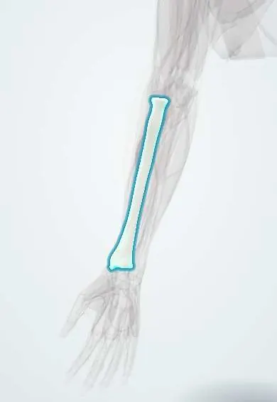
- Radius
- Ulna
- Interosseous membrane
- Radial head cartilage
- Annular ligament
- Elbow capsule
- Wrist capsule
- Posterior interosseous nerve
- Anterior interosseous nerve
- Median nerve
- Radial nerve
- Ulnar nerve
- Pronator quadratus
- Supinator
Why it happens
What it is
Fractures of the shaft of the radius, the ulna or both bones. Because the two bones work as a ring joined at the proximal and distal radioulnar joints, a displaced fracture of one bone is often accompanied by a fracture or dislocation of the other. Named patterns include the both-bone fracture, the Monteggia fracture-dislocation (proximal ulna fracture with radial head dislocation), the Galeazzi fracture-dislocation (distal radial shaft fracture with distal radioulnar joint disruption) and the isolated ulnar 'nightstick' fracture.
Who gets it
Forearm fractures are among the most common fractures of childhood, mostly after falls on an outstretched hand during play and sport. In adults they usually follow high-energy trauma, such as motorcycle crashes, which are a leading cause of injury in Colombia, or a direct blow when a person raises the arm in defence during an assault.
Causes
- Fall on an outstretched hand
- Motor vehicle and motorcycle crashes
- Direct blow to the forearm raised in self-defence (nightstick fracture)
- Sports and falls from height
- Pathological fracture through weakened bone (rare)
Risk factors
- Childhood and adolescence (active play, playground equipment, trampolines)
- High-risk sports and motorcycle riding
- Interpersonal violence
- Osteoporosis in older adults
Pathophysiology
Bending, twisting or direct forces break the shaft. In children the soft, thick periosteum allows incomplete patterns such as torus (buckle) and greenstick fractures, which may bend rather than break completely. Because the radius rotates around the ulna for pronation and supination, shortening or angulation of one bone forces dislocation at the proximal or distal radioulnar joint. Displaced fragments and swelling in the tight forearm compartments can injure the radial, median or ulnar nerves and raise compartment pressure.
Classification
- Both-bone shaft fracture (radius and ulna)
- Monteggia: proximal third ulna fracture with dislocation of the radial head (Bado types I–IV; type I anterior dislocation is most common)
- Galeazzi: distal third radial shaft fracture with dislocation of the distal radioulnar joint
- Isolated ulnar shaft (nightstick) fracture
- Paediatric patterns: torus (buckle), greenstick, complete and physeal (growth plate) fractures
- Open versus closed fracture
Symptoms and signs
- Pain and swelling of the forearm after injury
- Visible deformity or angulation
- Inability to rotate the forearm (pronation and supination)
- Tenderness at the elbow or wrist when a joint is dislocated
- Bruising and, in open fractures, a wound over the bone
Diagnosis
- Clinical
- Inspection for deformity and wounds, palpation of the whole forearm, elbow and wrist, and a documented neurovascular examination of the radial, median and ulnar nerves and the pulses before and after any manipulation.
- Tests
- No routine laboratory tests for isolated injuries; pre-operative blood tests as needed.
- Imaging
- Anteroposterior and lateral radiographs of the whole forearm that include the elbow and the wrist. On every view a line through the radial shaft and head should point to the capitellum; if not, the radial head is dislocated. CT is rarely needed.
Differential diagnosis
- Distal radius fracture (Colles or Smith)
- Supracondylar humerus fracture in children
- Radial head subluxation (pulled elbow) in toddlers
- Isolated elbow dislocation
- Soft-tissue contusion without fracture
Goal of treatment
Restore length, alignment and the rotational relationship of both bones so that the forearm regains full pronation and supination, while preventing compartment syndrome and nerve injury.
Treatment
- Acute
- Splinting in the position found, elevation, analgesia, removal of rings, neurovascular checks; antibiotics, tetanus prophylaxis and urgent debridement for open fractures.
- Conservative
- Torus fractures in children: a short splint or soft cast. Many displaced paediatric fractures: closed reduction under sedation and an above-elbow cast, accepting some angulation because of remodelling potential.
- Surgical
- Adults with displaced shaft fractures, and all Monteggia and Galeazzi injuries in adults: open reduction and internal fixation with plates and screws, which usually reduces the dislocated joint. Older children with unstable fractures may need flexible intramedullary nails.
- Follow-up
- Nursing priorities: hourly neurovascular observations of the hand in the first 24 hours, elevation, watching for escalating pain or tight cast, cast care education, and splitting the cast if circulation is compromised.
- Rehabilitation
- Early finger movement, then range-of-motion exercises for the elbow, wrist and forearm rotation after immobilisation.
Complications
- Compartment syndrome and Volkmann ischaemic contracture
- Loss of forearm rotation from malunion or radioulnar synostosis
- Missed radial head dislocation (chronic Monteggia lesion)
- Posterior interosseous nerve palsy
- Nonunion, refracture after plate removal, and infection after open fractures
Prognosis
Children generally heal quickly and remodel well, with excellent function. Adults treated with anatomical fixation usually regain good rotation; outcomes are worse when a Monteggia or Galeazzi dislocation is missed or when the fracture is open or comminuted.
Prevention
- Wrist guards and supervision for skating, playground and trampoline activities
- Motorcycle and road safety measures
- Fall prevention and osteoporosis treatment in older adults
Test yourself
Think of the answer, then tap the question to see it.
What injury defines a Monteggia fracture-dislocation?
A fracture of the proximal third of the ulna with dislocation of the radial head.
What injury defines a Galeazzi fracture-dislocation?
A fracture of the distal third of the radial shaft with dislocation of the distal radioulnar joint.
Which nerve is most often injured in a Monteggia fracture, and what deficit results?
The posterior interosseous nerve, causing weakness of finger and thumb extension without sensory loss.
Why must forearm radiographs include the elbow and the wrist?
To detect an associated dislocation of the radial head or of the distal radioulnar joint.
How are displaced adult forearm shaft fractures usually treated?
Open reduction and internal fixation with plates and screws.
Related conditions
- Distal radius fracture (Colles)
- Supracondylar humerus fracture
- Acute compartment syndrome
- Volkmann ischaemic contracture
- Open fracture
- Elbow dislocation
- Radial head subluxation (pulled elbow)
Educational information, not medical advice. For any symptom, see a health professional.
Sources: Forearm Fractures in Children - OrthoInfo - AAOS (American Academy of Orthopaedic Surgeons (OrthoInfo), 2025).
Spotted a mistake?We are always improving for the community. Tell us what is wrong and our team will review it.Report a mistake
Representative model, for educational purposes only.