
Pectus excavatum and pectus carinatum
The breastbone sinks in (funnel chest) or sticks out (pigeon chest). Most cases are mild, but a deep one can press on the heart; both can signal Marfan.
Quick facts
- Key signs
- A visible sunken or protruding chest · Breathlessness
- Diagnosis
- Low-dose CT or MRI
Red flags ↓
No doses: the clinician sets them by the current guideline.
What it is
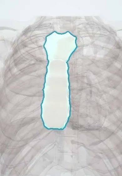
Pectus excavatum is a depression of the sternum and the lower costal cartilages into the chest, and pectus carinatum is a forward protrusion of the sternum. Both come from abnormal growth of the costal cartilages, usually worsen during the adolescent growth spurt, and range from a cosmetic difference to, in severe excavatum, compression of the heart and reduced exercise capacity.
Who gets it
Pectus excavatum is the commonest chest wall deformity, affecting about 1 in 300 to 1 in 1,000 children, three to five times more often boys. Pectus carinatum is several times less common, also mostly in boys, and usually appears or worsens at puberty. A family history is common. Both are seen in Marfan syndrome and other connective tissue disorders, which should be looked for in every patient.
Causes
- Unbalanced overgrowth of the costal cartilages, pushing the sternum in or out (the exact cause is unknown)
- Familial tendency in many cases
- Connective tissue disorders: Marfan syndrome, Ehlers–Danlos syndrome, homocystinuria
- Genetic syndromes: Noonan syndrome (often a combined upper carinatum and lower excavatum), Poland syndrome
- Acquired carinatum after median sternotomy in infancy or with severe chronic lung disease; rickets causes a different, rounded chest deformity
Risk factors
- Male sex
- Family history of chest wall deformity
- Tall, thin build with long limbs (think of Marfan syndrome)
- Scoliosis
- Rapid adolescent growth
Pathophysiology
Costal cartilages overgrow → in excavatum they buckle backward, carrying the lower sternum toward the spine → the distance between sternum and spine falls → the heart is pushed to the left and the right ventricle and atrium are compressed → stroke volume cannot rise normally with exercise → breathlessness, fatigue and palpitations on exertion; the lungs have less room → a mild restrictive pattern. Mitral valve prolapse is more common, partly from distortion and partly from shared connective tissue disease. In carinatum the cartilages push the sternum forward → the chest is fixed in an inspiratory, barrel-like position → mostly a cosmetic and pain problem, with little effect on the heart. In both, the change in body image during adolescence often matters more to the patient than the physiology.
Classification
- Pectus excavatum: symmetric or asymmetric; localised (cup-shaped) or diffuse (saucer-shaped); severity by depth and by the Haller index on cross-sectional imaging
- Haller index: the chest's width divided by the distance from the back of the sternum to the front of the spine at the deepest point; a normal chest is about 2.5, and a higher number means a deeper funnel (above about 3.25 has traditionally been called severe)
- Pectus carinatum: chondrogladiolar (lower sternum, keel-shaped, the commonest) or chondromanubrial (upper sternum, 'pouter pigeon', rare)
- Mixed forms: protrusion on one side and depression on the other, or upper carinatum with lower excavatum
- Syndromic (Marfan, Ehlers–Danlos, Noonan) versus isolated
Symptoms and signs
- A visible sunken or protruding chest, often first noticed in infancy and becoming more obvious in the growth spurt
- Breathlessness, reduced stamina or chest discomfort on exercise (severe excavatum)
- Palpitations; sometimes a systolic murmur or click of mitral valve prolapse
- Chest wall pain over the deformity (more in carinatum)
- Rounded shoulders and a forward-head posture
- Self-consciousness, avoidance of sports or swimming, and low mood about appearance
Diagnosis
- Clinical
- Inspect the chest from the front and side standing, measure the depth of the depression or the height of the protrusion, and check whether a carinatum flattens with manual pressure (flexible cartilage predicts brace success). Examine the spine for scoliosis and look actively for Marfan features (arm span greater than height, wrist and thumb signs, joint laxity, eyes) and for Noonan features. Ask about exercise tolerance and how the patient feels about the appearance.
- Tests
- Electrocardiogram (displaced axis and incomplete right bundle branch block can occur). Pulmonary function tests and cardiopulmonary exercise testing in symptomatic or severe excavatum to measure restriction and exercise capacity. Genetic evaluation (for example FBN1) when Marfan or another syndrome is suspected.
- Imaging
- Echocardiography for right heart compression, mitral valve prolapse and the aortic root (essential if Marfan is possible). CT or MRI of the chest at the deepest point to measure the Haller index and correction index and see cardiac displacement when surgery is considered; low-dose CT or MRI is preferred in young patients. A lateral chest X-ray gives a simple estimate. 3D surface scanning can follow change without radiation.
Differential diagnosis
- Marfan syndrome and other connective tissue disorders (pectus as a feature)
- Rickets (rachitic rosary and Harrison groove, not a true pectus)
- Poland syndrome (missing pectoralis muscle and chest asymmetry)
- Costochondritis or other causes of chest wall pain
- Asthma or deconditioning as the cause of breathlessness
- Scoliosis causing apparent chest asymmetry
A child with an unusual chest: which is it?
| Condition | What you see | Main problem | First approach |
|---|---|---|---|
| Pectus excavatum | Sternum sunken, deepest at the lower end | Compression of the right heart and lungs in severe cases; body image | Assess severity (Haller index, echo, exercise test); vacuum bell or surgery (Nuss) |
| Pectus carinatum | Sternum protruding like a keel | Mostly appearance and local pain | Compression brace while the cartilage is flexible; surgery if it fails |
| Marfan syndrome | Pectus with tall stature, long limbs and fingers | Aortic root dilation and dissection | Echocardiogram of the aortic root and genetic evaluation before any chest surgery |
| Rickets | Beaded costochondral junctions, groove along the lower ribs | Vitamin D or calcium deficiency | Treat the deficiency; the chest improves |
| Costochondritis | Normal shape; tender costal cartilages | Chest wall pain | Reassurance and simple analgesia |
Goal of treatment
Decide whether the deformity is only cosmetic or also limits the heart and lungs, rule out Marfan and related conditions, correct it with the least invasive method that works (bracing or a vacuum bell while the chest is pliable, surgery when needed), and support the patient's body image.
Treatment
- Conservative
- Mild deformity: observation through growth, posture exercises, chest and back strengthening and aerobic exercise; psychological support where appearance causes distress.
- Procedure
- Pectus carinatum: a dynamic compression brace worn many hours a day, adjusted to the pressure needed to flatten the chest, is first-line in growing adolescents with flexible cartilage. Pectus excavatum: a vacuum bell (a suction cup over the depression) can lift the sternum gradually in selected mild to moderate cases.
- Surgical
- Pectus excavatum with symptoms, cardiac compression or severe index: the minimally invasive Nuss procedure, passing one or more curved steel bars behind the sternum under thoracoscopic view to push it out, with the bars removed after a few years; or the open Ravitch operation (removal of the abnormal cartilages and repositioning of the sternum) for complex, asymmetric, adult or recurrent cases.
- Surgical
- Pectus carinatum not corrected by bracing: open cartilage resection (modified Ravitch) or minimally invasive compression with a bar placed in front of the sternum.
- Follow-up
- Pain after Nuss surgery is significant; multimodal analgesia, epidural or intercostal nerve cryoablation are used. In Marfan syndrome, check the aortic root first, time repair near the end of growth because recurrence is more common, and coordinate if heart surgery may be needed later.
Complications
- Severe excavatum: reduced exercise capacity, right heart compression, arrhythmias
- Mitral valve prolapse
- Psychological distress, social withdrawal and avoidance of sport
- After Nuss: bar displacement, pneumothorax, pleural effusion, wound infection, metal allergy, rarely heart or major vessel injury
- After open repair: recurrence, chest wall stiffness (acquired asphyxiating chest if done too young)
- Brace: skin irritation and pressure sores
Common traps
- Pectus excavatum is always just cosmetic: a deep funnel can compress the right heart and reduce exercise capacity; assess with an exercise test and imaging
- A chest deformity in a tall thin teenager only needs the chest fixed: look for Marfan syndrome first, because the aortic root, not the sternum, is what threatens life
- Pectus carinatum needs an operation: most respond to a compression brace while the cartilage is still flexible
- A higher Haller index means a less severe deformity: it is width divided by depth, so the deeper the funnel, the higher the index
- The best time to operate is early childhood: early open repair can limit chest growth; most repairs are done in adolescence, near the growth spurt
Prognosis
Most mild cases need no treatment. Bracing corrects most flexible carinatum when patients wear the brace as prescribed. Nuss and Ravitch repairs give good or excellent results in most patients, with improved appearance, quality of life and, in severe excavatum, often better exercise capacity; recurrence is uncommon but more frequent in connective tissue disorders.
Prevention
- No known prevention
- Early detection and follow-up through the growth spurt so bracing can start while the cartilage is flexible
- Screen every patient for Marfan syndrome and refer for echocardiography when features are present
Test yourself
Think of the answer, then tap the question to see it.
A 15-year-old with a deep funnel chest tires early in football and has a Haller index of 4.5. Why is he breathless?
The sternum compresses the right ventricle and atrium, so stroke volume cannot rise normally with exercise, and the lungs have less room. Surgery (Nuss) is reasonable.
A tall 14-year-old with pectus excavatum has long fingers and a high-arched palate. What must be done before chest surgery?
Assess for Marfan syndrome: echocardiogram of the aortic root and genetic evaluation. Aortic disease is the main risk, and repair timing and technique change with Marfan.
A 13-year-old has a keel-shaped chest that flattens when pressed by hand. What is the first-line treatment and why does the timing matter?
A dynamic compression brace. While the costal cartilages are still flexible in adolescence the brace can remodel them; after growth ends they stiffen and bracing works less.
How is the Haller index measured and what does a high value mean?
On an axial CT or MRI slice at the deepest point, the internal width of the chest divided by the distance from sternum to spine. Higher means deeper; about 2.5 is normal.
Connections
Educational information, not medical advice. For any symptom, see a health professional.
Sources: Pectus Excavatum (StatPearls (NCBI Bookshelf), 2026); Pectus Carinatum (StatPearls (NCBI Bookshelf), 2023); Pectus excavatum: MedlinePlus Medical Encyclopedia (MedlinePlus (U.S. National Library of Medicine), 2025).
Spotted a mistake?We are always improving for the community. Tell us what is wrong and our team will review it.Report a mistake
Representative model, for educational purposes only.