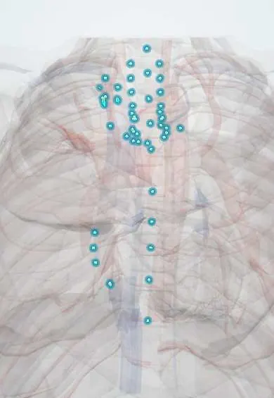
Mediastinitis (acute and fibrosing) !
Inflammation of the central chest compartment: a fast, deadly infection after surgery or a torn oesophagus, or slow scarring that squeezes vessels.
Updated 2026-11-01
Structures involved
Tap one to see what it is and open it in 3D.
- Mediastinal lymph nodes
- Esophagus
- Pericardium
- Sternum
- Superior vena cava
- Brachiocephalic veins
- Trachea
- Main bronchi
- Pulmonary arteries
- Pulmonary veins
- Thymus
- Pleura
- Phrenic nerves
- Recurrent laryngeal nerves
- Thoracic aorta
Why it happens
- Patterns of acute inflammation: serous, fibrinous, purulent, abscess, ulcer
- Fibrosis in organs: TGF-β and the myofibroblast
What it is
Inflammation of the mediastinum, the central chest space that holds the heart, great vessels, trachea, oesophagus and lymph nodes. Acute mediastinitis is a bacterial surgical emergency (after sternotomy, oesophageal perforation or a descending neck infection); fibrosing mediastinitis is slow post-infectious scarring, mostly after histoplasmosis, that narrows mediastinal vessels and airways.
Who gets it
Post-sternotomy mediastinitis complicates roughly 0.5 to 5% of median sternotomies and still carries a mortality around one in five. Mediastinitis after oesophageal rupture and descending necrotising mediastinitis from dental or deep neck infections are less common but more lethal when diagnosis is late. Fibrosing mediastinitis is rare and affects mainly young and middle-aged adults who lived in areas where Histoplasma is endemic; histoplasmosis occurs throughout Colombia and Latin America (caves, bat and bird droppings, poultry farms), and tuberculosis is a further cause in the region.
Causes
- Median sternotomy for cardiac surgery (deep sternal wound infection), mainly Staphylococcus aureus and coagulase-negative staphylococci
- Oesophageal perforation: endoscopy or dilation, forceful vomiting (Boerhaave syndrome), foreign bodies, trauma, anastomotic leak
- Descending necrotising mediastinitis: odontogenic infection, Ludwig angina, retropharyngeal or peritonsillar abscess tracking down the fascial planes (mixed oral flora including anaerobes)
- Penetrating chest trauma and airway perforation
- Fibrosing form: exaggerated immune response to past Histoplasma capsulatum infection, tuberculosis; less often sarcoidosis, silicosis, radiotherapy, IgG4-related disease or idiopathic
Risk factors
- Diabetes mellitus and poor perioperative glucose control
- Obesity, smoking and COPD
- Bilateral internal thoracic artery harvesting, re-operation, prolonged surgery or ventilation
- Oesophageal disease or recent endoscopic procedure
- Untreated dental infection or neck abscess
- Residence in a region endemic for histoplasmosis (fibrosing form)
Pathophysiology
The mediastinum is a loose connective-tissue space without anatomical barriers, continuous upward with the deep neck spaces (retropharyngeal and "danger" space) and lying directly behind the sternum and around the oesophagus. Bacteria entering from a sternal wound, a leaking oesophagus or the neck spread rapidly along these planes, causing cellulitis, abscess, tissue necrosis and sepsis; gastric contents leaking through an oesophageal tear add chemical injury. Infection can extend into the pleura (empyema), pericardium (purulent pericarditis, tamponade), sternum (osteomyelitis) and vessels (erosion, bleeding). In fibrosing mediastinitis, antigen from old granulomas in mediastinal lymph nodes drives a fibroinflammatory reaction that lays down dense collagen; this tissue compresses low-pressure, thin-walled structures first, such as the superior vena cava, pulmonary veins and arteries, main bronchi and oesophagus.
Classification
- Acute: post-sternotomy (deep sternal wound infection)
- Acute: after oesophageal perforation
- Acute: descending necrotising mediastinitis from the head and neck
- Chronic: granulomatous mediastinitis (enlarged caseous nodes, often silent)
- Chronic: fibrosing mediastinitis, focal (often calcified, post-infectious) or diffuse (often idiopathic or autoimmune)
Symptoms and signs
- Acute: high fever, chills, tachycardia and toxic appearance
- Acute: severe retrosternal or pleuritic chest pain, sometimes radiating to the neck or back
- After sternotomy: wound redness and purulent drainage, sternal instability or clicking, chest wall tenderness
- Subcutaneous emphysema in the neck or chest and a crunching sound with each heartbeat (Hamman sign) after oesophageal rupture
- Fibrosing: progressive breathlessness, cough, haemoptysis, recurrent pneumonia, dysphagia, and face and arm swelling
Diagnosis
- Clinical
- Suspect acute mediastinitis in any septic patient with chest pain after sternotomy, oesophageal instrumentation, vomiting or a deep neck infection; the window for effective treatment is short. Suspect fibrosing mediastinitis in a young adult with unexplained superior vena cava obstruction, pulmonary hypertension or recurrent haemoptysis, especially with past residence in a histoplasmosis area.
- Tests
- Blood count (leucocytosis), inflammatory markers, lactate and blood cultures. Gram stain and culture of wound drainage, pleural fluid or CT-guided mediastinal aspirate. In suspected rupture, a water-soluble contrast swallow or careful endoscopy locates the tear. For the fibrosing form: Histoplasma serology, tuberculin skin or interferon-gamma release test, and biopsy when lymphoma or another malignancy must be excluded.
- Imaging
- Chest X-ray may show a widened mediastinum, pneumomediastinum, air-fluid levels or pleural effusion. Contrast CT of neck and chest is the key test: fluid collections, gas bubbles, fat stranding, sternal dehiscence or an oesophageal leak. In fibrosing mediastinitis, CT shows an infiltrating, often calcified soft-tissue mass encasing and narrowing the superior vena cava, pulmonary vessels or bronchi; CT angiography and echocardiography assess vessel narrowing and pulmonary hypertension.
Differential diagnosis
- Acute coronary syndrome and aortic dissection
- Pulmonary embolism
- Pericarditis and pleural empyema without mediastinal spread
- Spontaneous pneumomediastinum without infection
- Mediastinal lymphoma or other mediastinal tumour (for the fibrosing form)
Goal of treatment
In acute mediastinitis: rapid source control and drainage with broad antibiotics and organ support to survive sepsis. In fibrosing mediastinitis: relieve obstruction of vessels and airways and treat complications, since the scar itself rarely regresses.
Treatment
- Medical
- Early broad-spectrum intravenous antibiotics covering staphylococci (including MRSA where prevalent), gram-negative bacilli and, after oesophageal or oral sources, anaerobes; antifungal cover in selected oesophageal leaks; adjusted to cultures.
- Surgical
- Urgent source control: sternal debridement, drainage and delayed closure or muscle flap reconstruction after sternotomy; repair or diversion of the oesophageal tear with mediastinal and pleural drainage; cervical and thoracic drainage for descending infection.
- Procedure
- Negative-pressure wound therapy for sternal wounds; endoscopic stenting or clipping of selected oesophageal leaks; for fibrosing disease, angioplasty and stents of the superior vena cava or pulmonary vessels and, rarely, airway stents.
- Medical
- Fibrosing form: antifungals do not reverse the fibrosis; corticosteroids or other immunosuppression are reserved for active inflammation or associated autoimmune or IgG4-related disease; symptomatic care for oedema, infections and haemoptysis.
- Acute
- Intensive care for sepsis: fluids, vasopressors, ventilation as needed, nil by mouth with enteral or parenteral nutrition after oesophageal injury, tight glucose control and nursing care of drains and wounds.
Complications
- Septic shock and multiorgan failure
- Empyema, purulent pericarditis and cardiac tamponade
- Sternal osteomyelitis and chronic sternal wound
- Erosion of great vessels with haemorrhage
- Fibrosing form: superior vena cava syndrome, pulmonary hypertension and right heart failure, bronchial stenosis with recurrent pneumonia, phrenic or recurrent laryngeal nerve palsy
Prognosis
Acute mediastinitis remains highly lethal: mortality after sternotomy exceeds about 20%, and after oesophageal perforation or descending necrotising infection it rises sharply when treatment is delayed beyond the first day. Fibrosing mediastinitis progresses slowly; disease limited to one side often allows a near-normal life expectancy, whereas bilateral involvement of pulmonary vessels or airways carries substantial mortality.
Prevention
- Perioperative antibiotic prophylaxis, skin preparation, glucose control and careful sternal closure in cardiac surgery
- Prompt treatment of dental and deep neck infections
- Careful technique during endoscopic dilation and early evaluation of chest pain after endoscopy
- Treating tuberculosis and limiting exposure to bat and bird droppings in histoplasmosis areas
Test yourself
Think of the answer, then tap the question to see it.
What are the three classic causes of acute mediastinitis?
Median sternotomy (deep sternal wound infection), oesophageal perforation and descending infection from the neck or mouth.
A patient vomits forcefully after heavy drinking and develops chest pain, fever and crepitus in the neck. What is the likely diagnosis and key test?
Boerhaave oesophageal rupture with mediastinitis; contrast CT and a water-soluble contrast swallow.
Which infection is the leading cause of fibrosing mediastinitis?
Histoplasmosis (Histoplasma capsulatum), followed by tuberculosis.
Which structures does fibrosing mediastinitis typically obstruct?
Low-pressure structures: superior vena cava, pulmonary veins and arteries, main bronchi and the oesophagus.
Why can a neck abscess cause mediastinitis?
The deep neck fascial spaces (retropharyngeal and danger space) open directly into the mediastinum, which has no barrier to spread.
Related conditions
- Oesophageal perforation (Boerhaave syndrome)
- Deep neck infection and Ludwig angina
- Histoplasmosis
- Pulmonary tuberculosis
- Sarcoidosis
- Superior vena cava syndrome
- Mediastinal masses (thymoma and anterior mediastinal mass)
- Parapneumonic effusion and empyema
- Acute pericarditis
- Cardiac tamponade
- Sepsis and septic shock
- IgG4-related disease
- Osteomyelitis
Educational information, not medical advice. For any symptom, see a health professional.
Sources: Mediastinitis: MedlinePlus Medical Encyclopedia (MedlinePlus Medical Encyclopedia (U.S. National Library of Medicine), 2024); Fibrosing Mediastinitis - Symptoms, Causes, Treatment | NORD (National Organization for Rare Disorders (NORD), 2025).
Spotted a mistake?We are always improving for the community. Tell us what is wrong and our team will review it.Report a mistake
Representative model, for educational purposes only.