
Complicated peptic ulcer: perforation and gastric outlet obstruction
A peptic ulcer can burn a hole through the gut wall or scar the stomach outlet shut. Perforation is a surgical emergency; obstruction causes vomiting.
What it is
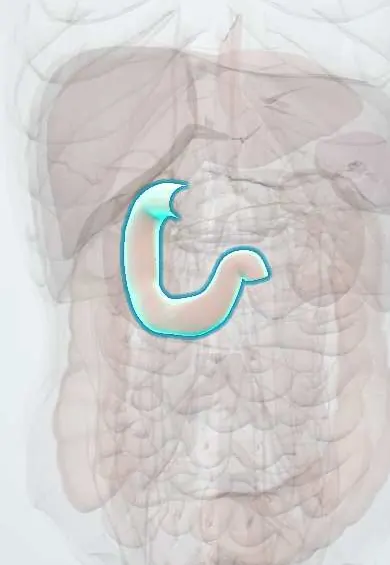
Two mechanical complications of peptic ulcer disease. Perforation is a full-thickness hole, usually in the anterior wall of the duodenal bulb or the stomach, that spills acid, food and later bacteria into the peritoneal cavity. Gastric outlet obstruction is narrowing of the pylorus or duodenum by oedema, spasm and scar from a chronic ulcer, so the stomach cannot empty.
Who gets it
Perforation affects a small minority of people with peptic ulcers but causes most ulcer deaths; it is commoner in older adults taking NSAIDs or aspirin and in smokers. Since effective acid suppression and Helicobacter pylori treatment, obstruction from benign ulcers has fallen, and in adults with outlet obstruction cancer must now be excluded first. In Colombia, high Helicobacter pylori prevalence, over-the-counter NSAID use and a high rate of gastric cancer make both points relevant.
Causes
- Helicobacter pylori infection
- NSAIDs, including low-dose aspirin, especially combined with corticosteroids, anticoagulants or antiplatelet drugs
- Smoking and heavy alcohol use
- Gastric cancer presenting as a perforated or obstructing ulcer
- Rarely, acid hypersecretion (Zollinger–Ellison syndrome) or cocaine and crack use
Risk factors
- Older age
- Long-term or high-intensity NSAID use without gastric protection
- Smoking
- Previous ulcer or ulcer complication
- Untreated Helicobacter pylori infection
- Severe physiological stress (critical illness, burns)
Pathophysiology
Acid and pepsin outmatch mucosal defence (Helicobacter pylori, NSAIDs) → the ulcer deepens through submucosa and muscle → anterior wall: it breaches the serosa → acid and food pour into the peritoneum (chemical peritonitis, sudden severe pain, board-like rigidity) → within hours bacteria follow (bacterial peritonitis), fluid shifts and sepsis set in → shock and organ failure, worse the longer the delay. Separately, a chronic pyloric or duodenal ulcer heals and breaks down repeatedly → oedema, spasm and fibrosis narrow the outlet → the stomach dilates and vomits retained food → loss of hydrochloric acid and volume → hypochloraemic, hypokalaemic metabolic alkalosis.
Classification
- Perforation: free (into the open peritoneum), sealed or contained (plugged by omentum or liver), or penetration into a neighbouring organ such as the pancreas
- By site: duodenal (most perforations, usually anterior bulb) or gastric (must be checked for cancer)
- Risk scores for perforation outcome: Boey score (shock, major medical illness, delay beyond about 24 hours) and PULP score
- Gastric outlet obstruction: benign (ulcer) or malignant (gastric, pancreatic or duodenal cancer)
Symptoms and signs
- Perforation: sudden, severe epigastric pain that spreads over the whole abdomen, often remembered to the minute
- Perforation: lying still, shallow breathing, pain in the shoulder tip from diaphragmatic irritation
- Perforation: rigid, board-like abdomen with guarding and absent bowel sounds; older or steroid-treated patients may show far less
- Obstruction: early fullness, bloating and vomiting of undigested food eaten hours or a day before, without bile
- Obstruction: weight loss, dehydration and a succussion splash over the stomach
Diagnosis
- Clinical
- Perforation: sudden epigastric pain followed by generalised peritonitis in a patient with ulcer history or NSAID use; ask about the time of onset. Obstruction: repeated vomiting of old food, weight loss and a succussion splash.
- Tests
- Full blood count, electrolytes and creatinine, lactate, blood gas, amylase or lipase (may be mildly raised in perforation), group and crossmatch and blood cultures. In obstruction: low chloride and potassium with metabolic alkalosis. Helicobacter pylori testing once the patient recovers, and biopsy of any gastric ulcer or stricture.
- Imaging
- Erect chest X-ray: free gas under the diaphragm in most, but not all, perforations. CT of the abdomen with contrast is more sensitive, shows small amounts of free gas and fluid and often the site. Water-soluble contrast study to show whether a perforation has sealed. Obstruction: CT shows a distended stomach and any mass; upper endoscopy after decompression shows the stricture and allows biopsy and dilatation.
Differential diagnosis
- Acute pancreatitis
- Oesophageal perforation (Boerhaave syndrome)
- Acute mesenteric ischaemia
- Acute cholecystitis or biliary colic
- Perforation of another hollow organ (colon diverticulum, appendix)
- Inferior myocardial infarction presenting as epigastric pain
- Malignant gastric outlet obstruction or gastroparesis (for obstruction)
Sudden severe upper abdominal pain: which one?
| Condition | Onset and pain | Bedside clue | Decisive test |
|---|---|---|---|
| Perforated peptic ulcer | Instant, severe epigastric pain that spreads to the whole abdomen | Board-like rigidity, lies still; NSAID or ulcer history | Free gas on erect chest X-ray or CT |
| Acute pancreatitis | Rapid onset, epigastric pain boring through to the back | Vomiting, prefers to lean forward; alcohol or gallstones | Lipase above three times normal; CT if unclear |
| Oesophageal perforation | Chest and upper abdominal pain after forceful vomiting | Neck crepitus (surgical emphysema), looks very ill | CT or contrast swallow showing leak and mediastinal gas |
| Acute mesenteric ischaemia | Severe pain out of proportion to a soft abdomen | Atrial fibrillation or vascular disease, older patient | CT angiography; lactate rises late |
| Acute cholecystitis | Builds over hours, right upper quadrant pain | Murphy sign, fever | Ultrasound: stones, thick wall, sonographic Murphy sign |
Goal of treatment
Perforation: resuscitate, control the source of contamination quickly and treat sepsis. Obstruction: decompress the stomach, correct fluid and electrolytes, exclude cancer and reopen the outlet. In both, cure the underlying ulcer disease so it does not return.
Treatment
- Acute
- Resuscitation: intravenous fluids, nil by mouth, nasogastric tube to empty the stomach, urinary catheter, analgesia, intravenous proton pump inhibitor and early broad-spectrum antibiotics.
- Surgical
- Perforation: laparoscopic or open closure of the hole, usually with a pedicle of omentum laid over it (omental patch repair), and washout of the peritoneal cavity. Perforated gastric ulcers are biopsied or excised because they may be cancers; large or difficult perforations may need resection.
- Conservative
- Selected stable patients with an early, sealed perforation (no contrast leak) may be managed without surgery: nasogastric suction, antibiotics and acid suppression with frequent surgical review, operating at once if they worsen. Not suitable for older patients or those in shock.
- Procedure
- Gastric outlet obstruction: nasogastric decompression, correction of chloride, potassium and volume, acid suppression, endoscopic biopsy and endoscopic balloon dilatation of a benign stricture.
- Surgical
- Obstruction that recurs after dilatation: a drainage operation such as gastrojejunostomy, or antrectomy; malignant obstruction is managed by stenting, bypass or resection according to the cancer.
- Medical
- After recovery: test for Helicobacter pylori and eradicate it, confirm eradication, stop NSAIDs where possible and continue acid suppression to heal the ulcer.
- Lifestyle
- Stop smoking, limit alcohol, and avoid over-the-counter anti-inflammatories without medical advice.
Complications
- Generalised peritonitis, septic shock and death
- Intra-abdominal abscess (subphrenic, pelvic)
- Leak from the repair
- Wound infection, pneumonia and respiratory failure after surgery
- Severe metabolic alkalosis, hypokalaemia and dehydration from obstruction
- Malnutrition and weight loss
- Missed gastric cancer
Common traps
- No free gas on the chest X-ray rules out a perforation: a sizeable minority of perforations show none on an erect film; with a convincing story, get a CT
- A perforation always follows years of ulcer pain: many patients, especially older NSAID or aspirin users, had no dyspepsia before the hole appeared
- The patch repair cures the ulcer: it only closes the hole; without Helicobacter pylori eradication and stopping NSAIDs, the ulcer and its complications come back
- A raised amylase settles it as pancreatitis: perforated ulcers can raise amylase too; free gas or a CT finds the perforation
- Vomiting old food in an adult with ulcer history is just a bad ulcer flare: it means outlet obstruction, and cancer must be excluded by endoscopic biopsy before calling it benign
Prognosis
Most patients with early surgery recover well, but mortality from perforation remains substantial and rises steeply with age, shock, other illness and delay to surgery. Benign outlet obstruction usually responds to dilatation or surgery; recurrence of either complication is uncommon once Helicobacter pylori is eradicated and NSAIDs are stopped.
Prevention
- Test for and eradicate Helicobacter pylori in people with ulcers
- Avoid unnecessary NSAIDs; use gastric protection (a proton pump inhibitor) in high-risk users
- Stop smoking
- Biopsy and follow gastric ulcers until healed
Test yourself
Think of the answer, then tap the question to see it.
A 72-year-old taking ibuprofen for knee pain has sudden severe epigastric pain, a rigid abdomen and gas under the right hemidiaphragm. What happened and what are the first steps?
A perforated peptic ulcer with peritonitis. Resuscitate, nil by mouth, nasogastric tube, intravenous proton pump inhibitor, broad-spectrum antibiotics and analgesia, and call the surgeon for omental patch repair.
Why do anterior duodenal ulcers tend to perforate while posterior ones tend to bleed?
The anterior wall faces the open peritoneal cavity, so a hole leaks into it; the posterior wall lies on the gastroduodenal artery and the pancreas, so erosion bleeds or penetrates instead.
A man with years of ulcer pain vomits large amounts of food eaten the day before. Which acid-base disorder do you expect, and why?
Hypochloraemic, hypokalaemic metabolic alkalosis: he loses hydrochloric acid in the vomit, and volume depletion makes the kidney keep sodium while losing potassium and hydrogen ions.
When can a perforated ulcer be managed without an operation?
In a young, stable patient seen early whose perforation has sealed (no leak on a contrast study), under close surgical review, operating at once if the patient worsens.
Why is a perforated gastric ulcer biopsied or excised at surgery?
Because a gastric cancer can perforate and look exactly like a benign ulcer; missing it delays cancer treatment.
Why it happens
In clinical skills
- Bowel perforation after a procedure: recognising it early
- When to call the surgeon: red flags, urgency and a good surgical referral
Connections
In physiology1
Educational information, not medical advice. For any symptom, see a health professional.
Sources: Peptic Ulcer Disease (StatPearls (NCBI Bookshelf), 2023); Duodenal Ulcer (StatPearls (NCBI Bookshelf), 2023); Gastric Outlet Obstruction (StatPearls (NCBI Bookshelf), 2025); Peptic Ulcer (MedlinePlus (U.S. National Library of Medicine), 2025).
Spotted a mistake?We are always improving for the community. Tell us what is wrong and our team will review it.Report a mistake
Representative model, for educational purposes only.