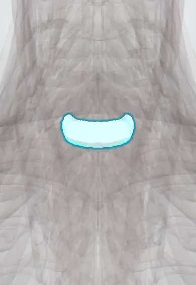
C7–T1 disc !
The C7-T1 disc is the cushion between C7 and T1, where the neck meets the upper back; it absorbs load and allows movement.
Official name: C7-T1
Opens in the Durgical app, no sign-up needed.
What it is
The cushion between C7 and T1: a tough outer ring of fibres (annulus fibrosus) around a soft centre.
What it does
Absorbs load and lets the neck bend and turn. C5-C6 is the most mobile and most worn neck level.
Key facts
- Where
- Between C7 and T1 bodies; cervicothoracic junction, below the vertebra prominens
- Attaches
- Annulus fibrosus anchored to the endplates and ring apophyses of C7 and T1; anterior and posterior longitudinal ligaments
- Function
- Absorbs and spreads load; posterolateral herniation usually compresses the C8 root
Relations
- In front
- Longus colli, esophagus and trachea entering the thorax; the recurrent laryngeal nerves in the tracheoesophageal grooves; on the left the thoracic duct arches nearby
- Behind
- Posterior longitudinal ligament, the dural sac and the spinal cord at the junction of neck and thorax
- Lateral
- The C8 root leaving through the C7–T1 foramen; further out, the sympathetic trunk (stellate ganglion) in front of the C7 transverse process and the neck of the first rib, and the dome of the pleura
- Below
- The T1 vertebra, the first thoracic one, which carries the head of the first rib
How to find it
From behind, between the vertebra prominens (C7 vertebra), usually the most prominent spinous process at the base of the neck, and the T1 spinous process just below; T1 is sometimes the more prominent of the two.
Examination
- Grip and finger flexion against resistance, with pinprick on the inner forearm: weakness plus forearm numbness points to the C8 root rather than the ulnar nerve
On imaging
On a plain lateral X-ray the shoulders often hide C7–T1; a swimmer's view (one arm raised) or CT shows the cervicothoracic junction. MRI levels here are counted from C2 down on a sagittal image.
Common problems
- Herniation pressing on the C6 nerve root: neck pain and pain, numbness or weakness down the arm into the thumb side of the hand.
- Degeneration with bone spurs, which can narrow the canal and press on the spinal cord (myelopathy: clumsy hands, unsteady walking).
How it is diagnosed
Exam of arm strength, reflexes and feeling, plus signs of cord pressure. MRI shows the disc, nerve roots and cord.
Treatment
- Most arm pain from a disc improves with time, physical therapy, medicine and sometimes an injection.
- Lasting arm pain, weakness or cord pressure: anterior cervical discectomy and fusion (ACDF), or disc replacement in selected patients.
Clinical notes
- Some surgeons prefer a left-sided anterior approach at the lowest cervical levels because the right recurrent laryngeal nerve takes a shorter, more oblique course around the subclavian artery; on the left the thoracic duct is the structure to protect
- In a short neck, the manubrium can block the anterior route to C7–T1
- After trauma the shoulders often hide C7–T1 on the lateral X-ray; the neck is not cleared until this level is seen, usually on CT, since injuries here are a classic miss
Anatomical variants
- Cervical rib: an extra rib, or a long fibrous band, from the C7 transverse process; uncommon, usually silent, but it can press on the lower brachial plexus trunk and imitate a C8 root lesion
Development
Every cervical transverse process holds a small costal element that normally stays fused to it. When the one on C7 grows on its own and forms a joint with the vertebra, it becomes a cervical rib.
Test yourself
Think of the answer, then tap the question to see it.
What is the clinical picture of the C8 root compressed by a C7–T1 disc herniation?
Weak finger flexion, grip and small hand muscles, numbness of the little and ring fingers and inner forearm; no standard reflex.
How is C8 radiculopathy from a C7–T1 disc told apart from ulnar nerve entrapment at the elbow?
Loss of feeling on the inner forearm (C8–T1) favours the root, since an ulnar lesion at the elbow spares it.
Why do some surgeons prefer a left-sided anterior approach to the C7–T1 disc?
The right recurrent laryngeal nerve takes a shorter, more oblique course around the subclavian artery; on the left the thoracic duct is the structure to protect.
Educational information, not medical advice. For any symptom, see a health professional.
Its names
- Official Latin (TA2)
- discus intervertebralis C7-T1 TA2 1684
- Official Spanish (DeCS)
- Disco C7-T1 DeCS D007403
- English (TA2)
- C7-T1
- Also called
- fibrous ring · fibrocartilago intervertebralis
Related
Sources: Terminologia Anatomica (TA2); DeCS; Anatomy, Back, Intervertebral Discs; Anatomy of the Human Body (20th ed.): Osteology Introduction (Lea & Febiger (Gray's Anatomy, 1918), 1918) · adapted, Public domain.
Spotted a mistake?We are always improving for the community. Tell us what is wrong and our team will review it.Report a mistake
Representative model, for educational purposes only.