
Mesenteric and omental disorders: the appendicitis mimics (mesenteric adenitis, omental infarction, mesenteric cysts, sclerosing mesenteritis)
Inflamed lymph nodes, a dead patch of fat or a cyst in the gut's fatty apron can hurt exactly like appendicitis. Most settle without surgery.
What it is
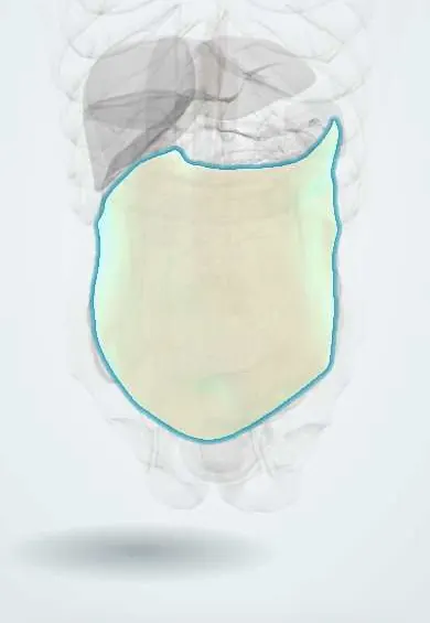
Disorders of the mesentery (the fold of peritoneum and fat carrying vessels and lymph nodes to the bowel) and greater omentum: mesenteric adenitis (inflamed mesenteric nodes), omental infarction (death of a segment of omental fat), mesenteric cysts and sclerosing mesenteritis (chronic inflammation and scarring of mesenteric fat).
Who gets it
Mesenteric adenitis is one of the commonest findings in children and adolescents operated on or scanned for suspected appendicitis. Omental infarction is uncommon, seen mostly in overweight adults and some obese children. Mesenteric cysts are rare, many found in children. Sclerosing mesenteritis is found in a small percentage of abdominal CT scans, mostly in men over 50, and usually causes no symptoms.
Causes
- Mesenteric adenitis: viral infection (often after an upper respiratory infection) or bacterial enteritis, classically Yersinia enterocolitica, also Campylobacter and Salmonella; secondary adenitis accompanies appendicitis, Crohn disease or lymphoma
- Omental infarction: torsion of a mobile segment of omentum or thrombosis of its fragile right-sided vessels; obesity, recent abdominal surgery, trauma and strenuous exercise predispose
- Mesenteric cysts: lymphatic malformations (cystic lymphangioma), enteric duplication cysts and mesothelial or dermoid cysts
- Sclerosing mesenteritis: cause unknown; associated with previous abdominal surgery or trauma, autoimmune disease, IgG4-related disease and malignancy, especially lymphoma
Risk factors
- Childhood and adolescence, recent viral illness (mesenteric adenitis)
- Obesity (omental infarction)
- Recent abdominal surgery or blunt trauma
- Male sex and age over 50 (sclerosing mesenteritis)
- Known or suspected lymphoma or other cancer
Pathophysiology
Mesenteric adenitis: organisms reach the terminal ileum → drain to the ileocolic lymph nodes in the right lower quadrant → the nodes swell and stretch the overlying peritoneum → right lower quadrant pain that copies appendicitis. Omental infarction: the right edge of the greater omentum is long, mobile and has a thin blood supply → it twists or its veins clot → the fat infarcts and inflames the adjacent parietal peritoneum → sharp, well-localised pain, usually on the right, with little systemic upset. Mesenteric cysts: a lymphatic channel fails to connect and fills with chyle or serous fluid → a mobile, slowly growing mass → pain only when it twists, bleeds, becomes infected or compresses the bowel. Sclerosing mesenteritis: chronic inflammation of mesenteric fat (panniculitis) → fat necrosis → fibrosis that can retract the mesentery, kink the bowel and narrow mesenteric vessels.
Classification
- Mesenteric adenitis: primary (no other cause found) or secondary (to appendicitis, Crohn disease, infection or lymphoma)
- Omental infarction: primary (spontaneous) or secondary (torsion around adhesions, a hernia or a tumour, or after surgery)
- Mesenteric cysts: by origin, lymphatic (lymphangioma), enteric, mesothelial, urogenital or dermoid; simple or complicated (torsion, haemorrhage, infection, rupture)
- Sclerosing mesenteritis: a spectrum from mesenteric panniculitis (inflammation and fat necrosis) to retractile mesenteritis (fibrosis)
Symptoms and signs
- Right lower quadrant or periumbilical pain, usually less severe and less focal in mesenteric adenitis than in appendicitis
- Mesenteric adenitis: fever, recent sore throat or cold, sometimes diarrhoea; tenderness may shift when the child lies on the side
- Omental infarction: sharp, constant right-sided pain over days in a patient who looks well, often without anorexia, vomiting or high fever
- Mesenteric cyst: painless abdominal swelling or a mobile mass; acute pain if it twists or bleeds
- Sclerosing mesenteritis: often none; otherwise recurrent abdominal pain, bloating, weight loss or a vague central mass
Diagnosis
- Clinical
- Approach as possible appendicitis first: history of a preceding viral illness, the sequence of pain, anorexia and vomiting, the site and severity of tenderness and peritonism. These diagnoses are usually made only once imaging shows a normal appendix. Re-examine within hours when unsure.
- Tests
- Full blood count and C-reactive protein (often mildly raised in adenitis and omental infarction), urinalysis and a pregnancy test in women of reproductive age. Stool culture if there is diarrhoea (Yersinia). In sclerosing mesenteritis or suspicious nodes: lactate dehydrogenase and tissue biopsy to exclude lymphoma; IgG4 levels when relevant.
- Imaging
- Ultrasound first in children and thin patients: a cluster of enlarged, oval mesenteric nodes in the right lower quadrant with a normal, compressible appendix suggests mesenteric adenitis. CT: omental infarction appears as a large, ill-defined fatty mass with stranding between the anterior abdominal wall and the colon, sometimes with a whirl of vessels; a mesenteric cyst as a thin-walled fluid-filled mass; sclerosing mesenteritis as hazy, denser mesenteric fat (misty mesentery) around the vessels, with a preserved fat halo around them and a thin surrounding pseudocapsule.
Differential diagnosis
- Acute appendicitis
- Epiploic appendagitis
- Meckel diverticulitis
- Terminal ileitis (Crohn disease, Yersinia, tuberculosis)
- Intussusception in young children
- Ovarian torsion, ruptured ovarian cyst or ectopic pregnancy
- Right-sided diverticulitis
- Lymphoma, carcinoid (neuroendocrine) tumour or desmoid tumour of the mesentery
Right lower quadrant pain: appendicitis or a mimic?
| Condition | Typical patient | Bedside clue | Imaging key | Usual management |
|---|---|---|---|---|
| Acute appendicitis | Child or young adult | Pain moves from the umbilicus to the right iliac fossa; anorexia, vomiting, peritonism | Enlarged, non-compressible appendix with surrounding fat stranding | Appendicectomy; antibiotics alone in selected uncomplicated cases |
| Mesenteric adenitis | Child or teenager after a cold or sore throat | Fever early, milder and less focal tenderness, often other enlarged nodes | Cluster of enlarged mesenteric nodes; normal appendix | Analgesia and observation; settles in days |
| Omental infarction | Overweight adult, sometimes an obese child | Sharp focal right-sided pain for days, looks well, little fever or vomiting | Large ill-defined fatty mass under the abdominal wall, often over 5 cm | Analgesia and observation; laparoscopic removal if not settling |
| Epiploic appendagitis | Adult, often overweight | Very focal pain, often in the left lower quadrant, little systemic upset | Small oval fatty lesion beside the colon with a bright rim and central dot | Anti-inflammatory analgesia; no surgery |
| Complicated mesenteric cyst | Child or young adult with a known or unnoticed mass | Mobile abdominal mass, acute pain if it twists or bleeds | Thin-walled fluid-filled cyst in the mesentery | Complete surgical excision |
Goal of treatment
Exclude appendicitis and other surgical emergencies, relieve pain, avoid an unnecessary operation when the diagnosis is benign and self-limiting, and remove or biopsy lesions that can obstruct, complicate or hide a malignancy.
Treatment
- Conservative
- Mesenteric adenitis: analgesia, fluids and observation with re-examination; symptoms usually settle within days. Antibiotics only for a proven bacterial cause with severe illness.
- Conservative
- Omental infarction and epiploic appendagitis: analgesia (including anti-inflammatory drugs) and observation once the CT diagnosis is clear; most resolve without surgery.
- Surgical
- Laparoscopic resection of the infarcted omentum when pain persists or worsens, when an abscess forms or when appendicitis cannot be excluded; it also shortens recovery for some patients.
- Surgical
- Mesenteric cyst: complete excision, usually laparoscopic, with a segment of bowel when the cyst shares its blood supply; drainage or partial removal alone tends to recur.
- Medical
- Symptomatic sclerosing mesenteritis: anti-inflammatory and immunomodulating treatment such as corticosteroids, sometimes tamoxifen or other agents; asymptomatic incidental cases need no treatment but follow-up imaging to exclude an underlying tumour.
- Surgical
- Surgery in sclerosing mesenteritis is reserved for biopsy of a suspicious mass and for bowel obstruction; the fibrosis usually cannot be resected.
Complications
- Missed appendicitis progressing to perforation
- Unnecessary appendicectomy (negative appendicectomy)
- Abscess or adhesions after omental infarction
- Torsion, volvulus, haemorrhage, infection or rupture of a mesenteric cyst
- Bowel obstruction or mesenteric ischaemia from retractile mesenteritis
- Missed lymphoma or other mesenteric tumour
Common traps
- Enlarged mesenteric nodes on ultrasound mean mesenteric adenitis: nodes also enlarge with appendicitis; the diagnosis needs a clearly normal appendix
- Focal right-sided pain with a raised CRP always needs an operation: a well-looking overweight adult with an omental infarct on CT usually settles with analgesia
- Mesenteric adenitis is a diagnosis to make on clinical grounds alone: without imaging or a period of observation it is a label that hides missed appendicitis
- A misty mesentery on CT is harmless and can be ignored: it is usually benign, but it can accompany lymphoma or other tumours, so look for a mass or nodes and follow it up
- A mesenteric cyst can simply be drained: incomplete removal tends to recur; complete excision is the treatment
Prognosis
Mesenteric adenitis, omental infarction and epiploic appendagitis are self-limiting and have an excellent outlook once correctly identified. Mesenteric cysts rarely recur after complete excision. Sclerosing mesenteritis is usually stable or regresses; the outlook depends mostly on whether an associated cancer is present.
Prevention
- No specific prevention; weight reduction may lower the risk of omental infarction
- Food and water hygiene reduces bacterial enteritis (Yersinia, Salmonella, Campylobacter)
- Imaging before surgery in stable patients with an uncertain diagnosis reduces negative appendicectomies
Test yourself
Think of the answer, then tap the question to see it.
A 9-year-old has right lower quadrant pain and fever a week after a cold. Ultrasound shows several enlarged nodes and a normal compressible appendix. What is the likely diagnosis and plan?
Mesenteric adenitis. Analgesia, fluids and observation with re-examination; return or rescan if pain worsens or peritonism appears.
Why does omental infarction usually affect the right side of the greater omentum?
The right edge is longer and more mobile, so it twists more easily, and its blood supply is thinner, so it infarcts when it twists or its veins clot.
A well-looking obese man has two days of sharp right-sided pain. CT shows a normal appendix and a large ill-defined fatty mass under the abdominal wall. What next?
Omental infarction: analgesia including anti-inflammatory drugs and observation; laparoscopic removal only if the pain persists, worsens or an abscess develops.
Why must a mesenteric mass seen with sclerosing mesenteritis be followed or biopsied?
Because the same CT appearance can accompany lymphoma, a neuroendocrine tumour or other cancer; a mass or bulky nodes should be sampled rather than assumed benign.
Connections
Educational information, not medical advice. For any symptom, see a health professional.
Sources: Mesenteric Adenitis (StatPearls (NCBI Bookshelf), 2023); Appendicitis (StatPearls (NCBI Bookshelf), 2024); Acute Abdomen (StatPearls (NCBI Bookshelf), 2025).
Spotted a mistake?We are always improving for the community. Tell us what is wrong and our team will review it.Report a mistake
Representative model, for educational purposes only.