
Non-ST-elevation acute coronary syndrome (NSTEMI and unstable angina)
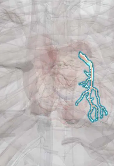
A clot on a cracked coronary plaque narrows the artery without fully shutting it. The heart is starved and the clot can still grow, so it is an emergency.
What it is
An acute coronary syndrome without persistent ST-segment elevation. If troponin rises and/or falls with at least one value above the 99th percentile, it is a non-ST-elevation myocardial infarction (NSTEMI); if troponin stays normal on serial testing despite rest or rapidly worsening angina, it is unstable angina.
Who gets it
NSTE-ACS is now more frequent than STEMI, and its patients are older, with more diabetes, kidney disease and previous coronary disease. Ischaemic heart disease is the leading cause of death worldwide and in Colombia. Women, older adults and people with diabetes more often present with breathlessness, fatigue or epigastric pain instead of classic chest pain.
Causes
- Rupture or erosion of an atherosclerotic plaque with a thrombus that narrows the artery or blocks it only on and off (the usual cause)
- Less often another acute coronary problem: spontaneous coronary artery dissection, coronary embolism or vasospasm
- Small platelet clots washed downstream (microembolisation), which cause patchy necrosis and the troponin rise
Risk factors
- Smoking, diabetes, high blood pressure and high LDL cholesterol
- Known coronary disease, previous MI, stent or bypass surgery
- Older age, male sex and a family history of early coronary disease
- Chronic kidney disease and peripheral or cerebrovascular disease
- Cocaine or other stimulant use
Pathophysiology
A plaque with a thin cap cracks or its surface erodes → platelets stick and activate, and thrombin builds a clot → the clot narrows the artery but leaves some flow (or blocks it only intermittently, or collaterals feed the territory) → the inner layer of the ventricle (subendocardium), furthest from the epicardial artery, is starved first → subendocardial ischaemia depresses the ST segment or inverts the T wave instead of elevating it → if ischaemia lasts long enough, subendocardial cells die and troponin rises (NSTEMI); if not, troponin stays normal (unstable angina) → the clot can grow to a full occlusion (STEMI) or shed emboli downstream, which is why antithrombotics are started at once and angiography follows at a speed set by risk.
Classification
- NSTEMI: ischaemic symptoms plus a rise and/or fall of troponin above the 99th percentile, without persistent ST elevation
- Unstable angina: angina at rest, new severe angina or crescendo angina, with normal troponin on serial high-sensitivity testing
- Risk group for angiography timing: very high risk (immediate), high risk (within 24 hours) and low risk (selective)
- Fifth Universal Definition (2026): an MI from a coronary plaque or other acute coronary problem is a primary MI; one caused by another acute illness through supply–demand imbalance is a secondary MI (formerly type 2)
Symptoms and signs
- Pressure, tightness or heaviness in the chest, at rest or with minimal effort, often lasting more than 20 minutes
- Pain spreading to the left arm, both arms, the jaw, neck or back
- Breathlessness, sweating, nausea or faintness
- Angina that is new, or more frequent, longer or easier to provoke than before (crescendo)
- Atypical presentations in women, older adults and people with diabetes: breathlessness, fatigue or epigastric pain alone
Diagnosis
- Clinical
- A 12-lead ECG within 10 minutes of first contact sorts the patient: persistent ST elevation (or a STEMI equivalent) goes down the STEMI pathway; everything else is assessed as possible NSTE-ACS with troponin and risk. Risk is then scored: GRACE (age, heart rate, systolic blood pressure, creatinine, Killip class, cardiac arrest at admission, ST deviation, raised troponin) predicts death, and a score above 140 means high risk; TIMI (age 65 or over, three or more coronary risk factors, known coronary stenosis, aspirin in the last week, two or more angina episodes in 24 hours, ST deviation, raised markers) is simpler. In the emergency department, HEART (History, ECG, Age, Risk factors, Troponin) helps decide who with chest pain can go home early; with high-sensitivity troponin pathways, the ECG and troponin carry most of its value.
- Tests
- ECG: ST depression, brief ST elevation or new T-wave inversion, but it may be normal; repeat it with every new pain, and record V7–V9 when V1–V3 show ST depression, because a posterior occlusion can hide as an «NSTEMI». High-sensitivity troponin by a validated 0/1-hour or 0/2-hour pathway: a very low value hours after pain began, or a low value that does not change, rules out MI; a high value or a clear rise rules in; values in between are observed and repeated. A rise and/or fall makes the injury acute; a stable raised value suggests chronic injury (kidney or heart disease). Creatinine, haemoglobin, glucose, lipids and electrolytes guide risk and drug choice.
- Imaging
- Echocardiography shows regional wall-motion abnormalities and the ejection fraction, and looks for the look-alikes (aortic dissection, tamponade, right ventricular strain from pulmonary embolism, takotsubo). Invasive coronary angiography finds and treats the culprit lesion. In low-risk patients once MI has been excluded, CT coronary angiography or stress imaging decides who needs invasive angiography.
Differential diagnosis
- STEMI or a STEMI equivalent (posterior MI, occlusion with left bundle branch block)
- Secondary MI (formerly type 2) and non-ischaemic myocardial injury: sepsis, tachyarrhythmia, anaemia, myocarditis, pulmonary embolism
- Aortic dissection
- Acute pericarditis
- Takotsubo syndrome
- Vasospastic angina
- Oesophageal spasm, reflux and chest wall pain
Chest pain or a raised troponin: which one is it?
| Condition | Mechanism | ECG | Troponin | What it needs |
|---|---|---|---|---|
| STEMI | Coronary artery usually fully blocked by a thrombus | Persistent ST elevation or an equivalent | Rises, but can still be normal in the first hours | Immediate reperfusion |
| NSTEMI | Thrombus on a plaque, artery partly or intermittently open | ST depression, T-wave inversion or normal | Rise and/or fall above the 99th percentile | Antithrombotics; angiography timed by risk |
| Unstable angina | Same thrombus, but no cell death | Like NSTEMI, or normal | Normal on serial testing | Antithrombotics; angiography or non-invasive testing by risk |
| Secondary MI (formerly type 2) | Supply–demand imbalance from another illness (tachyarrhythmia, anaemia, hypotension, hypoxia) | Often diffuse ST depression | Rise and/or fall | Treat the trigger, then look for underlying coronary disease |
| Non-ischaemic acute injury | Damage without ischaemia (myocarditis, sepsis, pulmonary embolism, trauma) | Depends on the cause | Rise and/or fall | Treat the cause, not as ACS |
| Chronic myocardial injury | Stable heart or kidney disease | Usual baseline | Raised but stable | Manage the underlying disease |
Goal of treatment
Stop the clot from growing, relieve ischaemia, and find and treat the culprit artery at a speed set by risk, while keeping bleeding as low as possible.
Treatment
- Acute
- Antiplatelet therapy: aspirin as soon as the diagnosis is likely, plus a P2Y12 inhibitor (ticagrelor, prasugrel or clopidogrel, chosen by bleeding risk and the planned strategy) as dual antiplatelet therapy. Routine P2Y12 pre-treatment before the coronary anatomy is known is no longer advised when early angiography is planned.
- Acute
- Parenteral anticoagulation (unfractionated or low-molecular-weight heparin, or fondaparinux) from diagnosis until angiography, chosen by kidney function, bleeding risk and timing. Fibrinolysis is not used in NSTE-ACS.
- Acute
- Anti-ischaemic therapy: nitrates for ongoing pain (not with hypotension, right ventricular infarction or recent phosphodiesterase-5 inhibitor use); an oral beta-blocker if there is no heart failure, shock or bradycardia; oxygen only when saturation is below 90%; opioids only for pain that nothing else relieves. NSAIDs are stopped.
- Procedure
- Angiography timed by risk: immediate (within about 2 hours) for very high risk (shock or haemodynamic instability, refractory pain, life-threatening arrhythmia or cardiac arrest, mechanical complications, acute heart failure from ischaemia, recurrent dynamic ST changes); early (within 24 hours) for high risk (confirmed NSTEMI, dynamic ST or T changes, brief ST elevation, GRACE above 140); selective for low risk, after non-invasive testing. The culprit lesion is treated by PCI or, in complex multivessel disease, bypass surgery.
- Medical
- Secondary prevention: dual antiplatelet therapy for a period set by ischaemic and bleeding risk, a high-intensity statin (with ezetimibe if LDL stays high), an ACE inhibitor or ARB with a weak ventricle, diabetes or hypertension, and a beta-blocker when the ejection fraction is reduced.
- Rehabilitation
- Cardiac rehabilitation with supervised exercise, education and psychological support.
- Lifestyle
- Stopping smoking completely, a heart-healthy diet, regular activity and weight control.
Complications
- Progression to full occlusion (STEMI) or recurrent ischaemia
- Heart failure and cardiogenic shock
- Ventricular arrhythmias and atrial fibrillation
- Bleeding from antithrombotics, at the access site or in the gut
- Contrast-associated acute kidney injury
- Mechanical complications (papillary muscle rupture, septal or free-wall rupture), rarer than in STEMI
Common traps
- No ST elevation means the artery is open: a circumflex or posterior occlusion often shows only ST depression in V1–V3; record V7–V9 and treat a true occlusion as a STEMI
- Every raised troponin is an NSTEMI: troponin marks myocardial injury of any cause; sepsis, a fast arrhythmia or anaemia cause secondary MI or non-ischaemic injury, which need the trigger treated rather than ACS drugs and urgent angiography
- NSTEMI should get thrombolysis like STEMI: fibrinolysis gives no benefit in NSTE-ACS and adds bleeding; the treatment is antiplatelets, an anticoagulant and angiography timed by risk
- A normal first troponin rules out MI: an early presenter needs a repeat at 1 or 2 hours by a validated pathway; only a very low value hours after pain began rules out on one sample
- A stable NSTEMI never needs urgent angiography: refractory pain, shock, heart failure or ventricular arrhythmia make it very high risk, and angiography is then done within about 2 hours
Prognosis
Death in hospital is lower than in STEMI, but over the following months it catches up, because patients are older and have more comorbidity. A high GRACE score, ongoing ischaemia, heart failure, kidney disease and diabetes mark the worst outcomes; timely angiography and secondary prevention improve them.
Prevention
- Control of smoking, blood pressure, diabetes and LDL cholesterol
- Secondary prevention drugs and cardiac rehabilitation after any acute coronary syndrome
- Seeking care at once for new or worsening chest pain
Test yourself
Think of the answer, then tap the question to see it.
Which test result separates NSTEMI from unstable angina?
Troponin. NSTEMI has a rise and/or fall above the 99th percentile; in unstable angina troponin stays normal on serial high-sensitivity testing.
Why does NSTE-ACS usually cause ST depression rather than ST elevation?
The artery is not fully blocked, so ischaemia is mostly subendocardial (the inner layer, furthest from the epicardial artery), which depresses ST; full occlusion causes transmural ischaemia, which elevates it.
A 67-year-old with NSTEMI has recurrent chest pain, hypotension and pulmonary oedema despite treatment. How soon is angiography done, and why?
Immediately, within about 2 hours: refractory pain, shock and acute heart failure mark very high risk, where ongoing ischaemia threatens life.
A septic patient with fast atrial fibrillation has a troponin that rises and falls. What is the likely diagnosis and the priority?
Secondary MI (formerly type 2) or non-ischaemic injury from supply–demand imbalance. Treat the sepsis and control the rate; ACS antithrombotics and urgent angiography are not given by default.
Which findings put an NSTE-ACS patient in the high-risk group for angiography within 24 hours?
A confirmed NSTEMI, dynamic ST or T-wave changes, brief ST elevation, or a GRACE score above 140.
Why it happens
In rehabilitation
Connections
Educational information, not medical advice. For any symptom, see a health professional.
Sources: Non–ST-Segment Elevation Myocardial Infarction (NSTEMI) (StatPearls (NCBI Bookshelf), 2023); Acute Coronary Syndrome (StatPearls (NCBI Bookshelf), 2026); Fifth Universal Definition of Myocardial Infarction (2026) (Global Heart (ESC/ACC/AHA/WHF Task Force), open access, 2026); Current Management of Non-ST-Segment Elevation Acute Coronary Syndrome (Biomedicines (open access, PMC), 2024); The HEART score has less utility with high sensitivity troponin (Frontiers in Cardiovascular Medicine (open access, PMC), 2026); Contemporary Challenges and Strategies for Diagnosing and Managing in Type 2 Myocardial Infarction: A Narrative Review (Journal of Personalized Medicine (open access, PMC), 2026).
Spotted a mistake?We are always improving for the community. Tell us what is wrong and our team will review it.Report a mistake
Representative model, for educational purposes only.