
Retained surgical items (gossypiboma)
A sponge, needle or instrument left inside after surgery is rare and preventable, but it can cause infection, obstruction or a fake tumour years later.
What it is
A retained surgical item is any object used during an operation and unintentionally left in the patient: soft items such as sponges, gauze and towels (a retained textile is called a gossypiboma or textiloma), and hard items such as needles, blades, instruments, wire and device fragments. It is considered a never event because it is preventable with reliable systems.
Who gets it
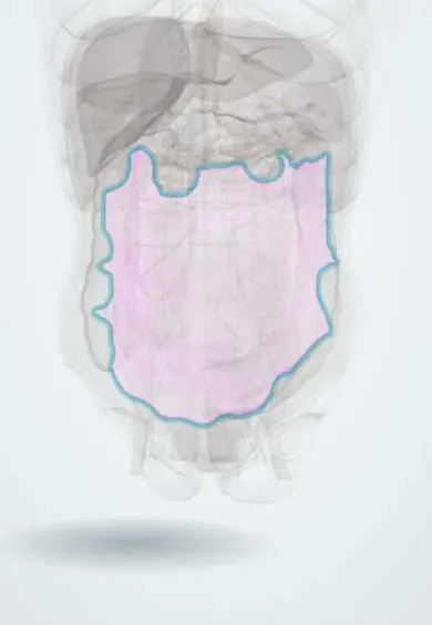
Estimates range from about 1 in 5,500 to 1 in 18,760 inpatient operations, and the true rate is probably higher because many cases surface late or are never reported. Sponges are the classic item, but hard items such as needles and instruments now make up a growing share. Retained items have been the most frequently reported sentinel event in hospital safety reporting systems. The abdomen and pelvis, including caesarean section and hysterectomy, are the commonest sites.
Causes
- Soft items: sponges, laparotomy pads, gauze, towels and packs (gossypiboma)
- Hard items: suture needles, blades, clamps, retractors, electrode tips, guidewires and fragments of devices
- Packs left on purpose in damage control surgery that are not all removed at the planned re-look
- Dressings and sponges from negative-pressure wound therapy left in the wound
Risk factors
- Emergency surgery
- An unplanned change in the operation, or unexpected findings
- High body mass index
- A count discrepancy, or no count at all
- Heavy blood loss, long operations, more than one procedure or more than one surgical team
- Change of nursing or surgical staff during the operation, and inexperienced or too few staff
- Communication failures within the team
Pathophysiology
The item sets off a foreign-body reaction → two paths follow. Fibrinous (aseptic) path: fibroblasts wall it off in a capsule → a firm, sometimes calcified mass with adhesions, silent for months or years → found as an incidental mass or mistaken for a tumour. Exudative path: bacteria and inflammation around the textile → abscess → the body tries to push it out through a fistula to the skin, or by erosion into the bowel, bladder or vagina → the item can migrate into the bowel lumen and cause obstruction, malabsorption or bleeding. Hard items, especially needles, may stay inert, migrate, or injure nearby structures.
Classification
- By item: soft (textiles) or hard (needles, instruments, fragments)
- By reaction: fibrinous (encapsulated mass) or exudative (abscess, fistula, migration)
- By timing: discovered during the operation (near miss), early after surgery, or late (months to years)
- Intentional packing left for a planned re-look is not a retained item unless it is forgotten
Symptoms and signs
- Often none for a long time
- Early: fever, pain and a collection after surgery that does not settle; sepsis
- Persistent wound discharge or a sinus that will not heal
- A palpable abdominal or pelvic mass
- Bowel obstruction, gastrointestinal bleeding or passage of fragments of gauze through the rectum after migration
- Urinary symptoms or vaginal discharge when the item erodes into the bladder or vagina
Diagnosis
- Clinical
- During surgery, a count that does not match means the item is in the patient until proven otherwise: stop, search the wound and the field, and image before closing if it is not found. Months or years later, suspect it in anyone with an unexplained mass, collection or fistula at a previous operative site, and ask for the full operative history.
- Tests
- Blood count and inflammatory markers for infection; cultures from collections and fistula discharge. Histology of a resected mass may be the first proof in a fibrinous gossypiboma mistaken for a tumour. Every count discrepancy and every retained item is reported through the institution's safety system.
- Imaging
- Intraoperative or postoperative plain radiography detects sponges with radio-opaque markers, but it misses a meaningful share of items (false negatives of about 10 to 25 percent in reports), and small needles are hard or impossible to see; the radiologist needs to know exactly what is missing. CT is the best test after surgery: a textile typically appears as a mass with a spongiform pattern of trapped gas bubbles, sometimes a wavy or whorled internal pattern, and a calcified rind in old cases. Ultrasound shows a cystic mass with a wavy echogenic structure inside, or a solid mass with strong acoustic shadowing. MRI helps when CT is unclear.
Differential diagnosis
- Postoperative abscess or haematoma without a foreign body
- Tumour or recurrence of cancer at the operative site
- Intentional surgical haemostatic material or a drain
- Bowel loops with gas and faeces mimicking a gas-containing mass
- Other causes of fistula: anastomotic leak, Crohn disease, radiation
Retained surgical item: how it shows itself
| Path | When | How it presents | Imaging clue |
|---|---|---|---|
| Exudative: abscess | Days to weeks | Fever, pain, a collection that does not settle | Collection containing a gas-filled spongiform mass |
| Fibrinous: encapsulated mass | Months to years | Silent, or a firm mass mistaken for a tumour | Mass with a whorled or wavy internal pattern, sometimes a calcified rim |
| Fistula or migration | Weeks to years | Discharge from the skin, obstruction, bleeding, gauze passed per rectum | Mass inside or tracking into the bowel, bladder or vagina |
| Hard item (needle, instrument) | Any time, often found on an X-ray | Often silent; pain or injury if it migrates | Metallic density; small needles may not show on plain X-ray |
Goal of treatment
Prevent retention with layered, team-based systems; when a count is wrong, find the item before the patient leaves the theatre; and when an item is found later, remove it safely, treat its complications, disclose it to the patient and learn from it without blame.
Treatment
- Acute
- Count discrepancy during surgery: tell the whole team, pause closure, search the wound and the field and the waste, and obtain an intraoperative X-ray of the operative area if the item is still missing; document the outcome.
- Surgical
- Removal of the retained item by laparotomy or laparoscopy, with drainage of any abscess, repair or resection of eroded bowel and closure of fistulas.
- Medical
- Antibiotics for associated infection, guided by cultures, alongside source control.
- Follow-up
- Open disclosure to the patient and family, reporting through the hospital safety system, and a review of the event that looks at the system rather than blaming one person.
Complications
- Abscess and sepsis
- Enterocutaneous and internal fistulas
- Bowel obstruction, perforation, bleeding and malabsorption after migration
- Unnecessary cancer workup or major resection for a pseudotumour
- Reoperation, longer hospital stay and higher costs
- Psychological harm, loss of trust and medicolegal claims
Common traps
- A correct count means nothing was left: in most reported cases the final count was recorded as correct; risk factors such as emergency surgery or a change of plan call for extra checks
- A negative X-ray rules out a retained item: plain films miss some marked sponges and many small needles; tell the radiologist exactly what is missing
- A gas-containing mass after surgery is always an abscess: a spongiform pattern of trapped gas inside a mass is the classic CT sign of a retained textile
- A new mass years after surgery must be cancer: a fibrinous gossypiboma can look like a tumour; the operative history and the CT pattern raise the question before a major resection
- Retained items are one careless person's fault: they come from system failures (emergencies, fatigue, hand-overs, poor communication), so prevention is built into the team's routine
Prognosis
Once removed, most patients recover, but the harm depends on how long the item stayed and what it eroded into. Many retained items are found only years after the operation. Because counts were recorded as correct in most reported cases, counting alone is not enough, which is why technology and imaging are added.
Prevention
- Standard counts of sponges, sharps and instruments before the operation, whenever items are added, before closing a cavity, at wound closure and at skin closure
- Only sponges with radio-opaque markers in body cavities
- A methodical wound sweep before closure
- Adjunct technology such as barcode or radiofrequency-tagged sponges, especially in high-risk operations
- Routine X-ray before closing in high-risk cases (emergencies, unexpected changes, high body mass index) and after any unresolved discrepancy
- Closed-loop communication and a surgical safety checklist, with every team member free to stop the closure
Test yourself
Think of the answer, then tap the question to see it.
At the end of an emergency laparotomy, one laparotomy pad is missing from the count. What happens next?
The team stops closure, searches the wound, field and waste, and if the pad is not found takes an intraoperative X-ray before closing, documenting the result.
Why are emergency operations, unplanned changes of procedure and a high body mass index risk factors for retained items?
They create rushed, complex conditions with many items in deep spaces, where counts are skipped or wrong and items hide easily.
A woman has a pelvic mass 3 years after a caesarean section. CT shows a mass with trapped gas bubbles and a wavy internal pattern. What should be suspected?
A retained surgical sponge (gossypiboma), which can mimic a tumour or an abscess; removal confirms it.
Why can a negative postoperative X-ray not fully exclude a missing needle?
Small needles are poorly visible or invisible on plain radiographs, and even marked sponges are sometimes missed.
Connections
Educational information, not medical advice. For any symptom, see a health professional.
Sources: Retained surgical sponges, needles and instruments (Annals of the Royal College of Surgeons of England (PMC), 2013); Risk factors and preventive strategies for unintentionally retained surgical sharps: a systematic review (Patient Safety in Surgery (PMC), 2021); A Pictorial Review of the Many Faces of Gossypiboma – Observations in 6 Cases (Polish Journal of Radiology (PMC), 2017); Retained Surgical Foreign Bodies after Surgery (Open Access Macedonian Journal of Medical Sciences (PMC), 2017).
Spotted a mistake?We are always improving for the community. Tell us what is wrong and our team will review it.Report a mistake
Representative model, for educational purposes only.