
Anastomotic leak
A hole where two ends of bowel were joined in surgery, letting gut contents escape. It is the complication surgeons fear most after bowel surgery.
What it is
A defect in the wall at a surgical join (anastomosis) of the gut that lets luminal contents pass from inside to outside the bowel. It may stay contained as an abscess or spread as peritonitis, mediastinitis or empyema, and usually shows itself between days 3 and 8 after surgery, sometimes later.
Who gets it
Leak rates vary with the site: lowest after small-bowel and ileocolic joins, higher after oesophageal and pancreatic anastomoses, and highest for low rectal anastomoses close to the anus, reaching well over 10% in some series. Leaks account for a large share of deaths after colorectal surgery and many unplanned reoperations.
Causes
- Poor blood supply to the bowel ends (ischaemia), the most important technical cause
- Tension on the join
- Technical failure of sutures or staples
- Poor healing host: malnutrition, steroids, prior radiotherapy, sepsis, smoking
- Contamination, emergency surgery and raised pressure inside the bowel (distal obstruction)
Risk factors
- Low rectal anastomosis (close to the anal verge), oesophageal anastomosis
- Male sex and a narrow pelvis, obesity
- Neoadjuvant chemoradiotherapy
- Malnutrition, low albumin, anaemia, diabetes
- Smoking, heavy alcohol use, long-term steroids or immunosuppression
- Emergency surgery, long operation, intraoperative blood loss and transfusion, high ASA class
Pathophysiology
Ischaemia, tension or poor host healing → the join fails to seal during the lag phase of healing (days 3–5) → a defect lets bowel contents and bacteria out → if adjacent tissue walls it off: localised abscess, sometimes later a fistula to skin, vagina or bladder → if not contained: generalised peritonitis (or mediastinitis and empyema after oesophagectomy) → sepsis, ileus and organ failure → late consequences: anastomotic stricture, poor bowel function and, in cancer surgery, higher local recurrence.
Classification
- Grade A (rectal leak, international definition): radiological or subclinical leak that does not change management
- Grade B: needs active treatment but no reoperation (antibiotics, percutaneous or transanal drainage)
- Grade C: needs reoperation
- By time: early (first days, often technical) versus late (after the first week, often host or ischaemia related)
- By extent: contained (abscess) versus free (generalised peritonitis, mediastinitis)
Symptoms and signs
- Fever and tachycardia, often the earliest signs; new atrial fibrillation, especially after oesophagectomy
- Abdominal or pelvic pain beyond what is expected, peritonism
- Ileus that does not resolve or returns after improvement
- Feculent, purulent, bilious or turbid fluid in a drain
- Confusion, low urine output, breathing difficulty
- Rectal pain or discharge (low rectal leak); neck wound redness or chest pain (oesophageal leak)
Diagnosis
- Clinical
- Suspect it in any patient who is not progressing as expected after an anastomosis: tachycardia, fever, pain, prolonged ileus, organ dysfunction or abnormal drain fluid. Early-warning scores used daily help catch the change.
- Tests
- CRP trend (a low and falling CRP around days 3–5 makes a leak unlikely; a high or rising one calls for imaging), full blood count, lactate, renal function, blood cultures; drain fluid appearance and, after pancreatic surgery, drain amylase.
- Imaging
- CT of the abdomen and pelvis with intravenous contrast plus water-soluble contrast given rectally (colorectal) or orally (upper gut): extravasation of contrast, gas and fluid around the join. After oesophagectomy, CT with oral contrast and endoscopy, which also shows the size of the defect and graft viability. Flexible endoscopy or a contrast enema for low rectal joins.
Differential diagnosis
- Intra-abdominal abscess without a leak
- Prolonged postoperative ileus
- Pneumonia or other hospital-acquired infection
- Pulmonary embolism
- Bleeding from the anastomosis or the operative field
- Mesenteric ischaemia of the conduit or bowel
Not recovering after a bowel resection: leak or something else?
| Problem | Typical timing | Key clue | CT finding | Usual move |
|---|---|---|---|---|
| Anastomotic leak | Days 3–8 | Tachycardia, pain, rising CRP, bowel contents in drain | Contrast escaping at the join, gas and fluid | Source control: drain, endoscopic therapy or reoperation |
| Intra-abdominal abscess | Days 5–10 | Swinging fever, localised pain or tenderness | Rim-enhancing collection, no contrast leak | Percutaneous drainage and antibiotics |
| Postoperative ileus | From day 1, lasting | Distension and vomiting, patient otherwise well, CRP falling | Dilated bowel without collection or leak | Nasogastric tube if vomiting, correct electrolytes, reduce opioids |
| Surgical site infection | Days 5–7 | Red, tender, discharging wound | Superficial collection only | Open and drain the wound |
Goal of treatment
Recognise the leak early, resuscitate, control the source with the least invasive method that works, and protect life first and the anastomosis second.
Treatment
- Acute
- Resuscitation and sepsis care: fluids, broad-spectrum antibiotics covering bowel flora, oxygen, and organ support in the ICU if needed.
- Procedure
- Contained leak with an abscess: CT- or ultrasound-guided percutaneous drainage, or transanal drainage for a low rectal leak; endoscopic vacuum therapy for rectal or oesophageal leaks; covered stents or clips for selected oesophageal or gastric leaks.
- Surgical
- Generalised peritonitis or an unstable patient: reoperation, washout and usually taking the join apart with an end stoma (for example a Hartmann procedure), or repair with a proximal diverting stoma in selected cases.
- Conservative
- Small, contained, clinically mild leaks (grade A or some grade B) in a patient who already has a diverting stoma may be managed with antibiotics, bowel rest and close observation.
- Lifestyle
- Nutrition support during recovery, enteral when the gut can be used (for example a feeding jejunostomy or tube beyond an oesophageal leak), parenteral when it cannot.
- Follow-up
- Nursing priorities: heart rate, temperature, blood pressure, urine output and pain every few hours, drain output and character, early-warning scores, and immediate escalation of a patient who stops progressing.
Complications
- Peritonitis, sepsis and death
- Pelvic or intra-abdominal abscess
- Fistula to skin, vagina or bladder
- Permanent stoma
- Anastomotic stricture and poor bowel function (low anterior resection syndrome)
- Higher local recurrence and worse survival after cancer surgery
Common traps
- No fever means no leak: tachycardia, new atrial fibrillation, confusion or a stalled recovery are often the first signs; temperature may stay normal
- A diverting stoma prevents leaks: it reduces their rate and especially their consequences, but the join can still leak; it changes management, usually allowing drainage instead of reoperation
- An abnormal patient on day 2 is too early for a leak: leaks peak around days 3 to 8, but technical failures can show within 48 hours; do not wait for the textbook day
- Prolonged ileus is just slow bowel: ileus that does not settle, or returns, is a classic presentation of a leak or abscess; check CRP and image
- A drain will always show the leak: drains are often blocked, misplaced or walled off; clean drain fluid does not rule out a leak
Prognosis
Mortality rises steeply with delayed diagnosis and with free rather than contained leaks. Many patients with a low rectal leak never have their stoma reversed, and bowel function and cancer outcomes are worse after a leak.
Prevention
- Careful technique: well-perfused ends, no tension, airtight join (leak test for left-sided joins); fluorescence angiography can confirm perfusion
- Temporary diverting ileostomy for low rectal anastomoses, which reduces the consequences and the rate of clinical leaks
- No anastomosis in unstable, contaminated or septic patients (end stoma or damage-control surgery instead)
- Correct malnutrition and anaemia, stop smoking and optimise diabetes before elective surgery
- Mechanical bowel preparation with oral antibiotics before elective colorectal surgery where protocols include it
Test yourself
Think of the answer, then tap the question to see it.
On day 4 after a low anterior resection a patient has a pulse of 120 and pelvic pain but a normal temperature. What is the next step?
Treat as a possible anastomotic leak: resuscitate, check CRP and lactate, start antibiotics if septic, and get CT with intravenous and rectal water-soluble contrast.
Why is an anastomosis most vulnerable around days 3 to 5?
In the early lag phase collagen breakdown exceeds new synthesis, so the join depends on sutures or staples just as bowel function returns and pressure inside rises.
Which three conditions does an anastomosis need in order to heal?
Good blood supply to both ends, no tension, and a patient able to heal (nourished, not septic, without heavily irradiated tissue or high-dose immunosuppression).
A patient with generalised peritonitis and septic shock from a colorectal leak: drainage or reoperation, and why?
Reoperation: free leakage with shock needs washout and usually taking down the join with an end stoma; percutaneous drainage suits contained collections in stable patients.
In clinical skills
Connections
Educational information, not medical advice. For any symptom, see a health professional.
Sources: Colorectal anastomotic leakage: a narrative review of definitions, grading systems, and consequences of leaks (Frontiers in Surgery (PMC), 2024); Colorectal anastomotic leakage: aspects of prevention, detection and treatment (World Journal of Gastroenterology (PMC), 2013); Anastomotic leakage after esophagectomy for esophageal cancer: definitions, diagnostics, and treatment (Diseases of the Esophagus (PMC), 2021).
Spotted a mistake?We are always improving for the community. Tell us what is wrong and our team will review it.Report a mistake
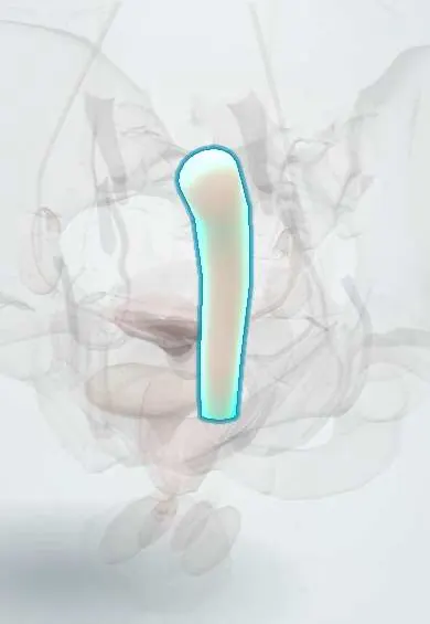
Representative model, for educational purposes only.