
Spinal cord and spinal canal tumours
Tumours can grow inside the spinal cord, beside it within its covering, or outside it in the bone. Where they sit predicts what they are and how they present.
Quick facts
- Key signs
- «Biologic» back pain at night · Mechanical pain worse
- Diagnosis
- Biopsy or surgical histology
Red flags ↓
No doses: the clinician sets them by the current guideline.
What it is
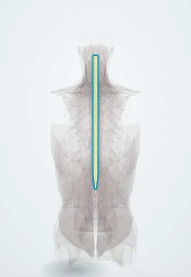
Primary or secondary tumours of the spinal cord, its coverings or the surrounding spine, grouped by compartment: intramedullary (inside the cord), intradural-extramedullary (inside the dura, outside the cord) and extradural (outside the dura, mostly metastases).
Who gets it
Uncommon. Intramedullary tumours make up about 2 to 4% of central nervous system tumours. Schwannomas, meningiomas and ependymomas together account for about four in five primary spinal tumours. Ependymoma is the most common intramedullary tumour in adults (about 60% of adult cases), while pilocytic astrocytoma is the most common in children (60 to 70%). Spinal meningiomas mainly affect women in their fifth decade and are most often thoracic. Most extradural tumours are metastases.
Causes
- Primary tumours of the cord's glial and ependymal cells: ependymoma, astrocytoma
- Tumours of the coverings and nerve roots: meningioma (from arachnoid cap cells), schwannoma and neurofibroma
- Vascular tumours: haemangioblastoma, sporadic or in von Hippel-Lindau disease
- Metastases to the spine from lung, breast, prostate, kidney and thyroid cancers; myeloma and lymphoma; primary bone tumours such as chordoma and sarcomas
Risk factors
- Neurofibromatosis type 2 (NF2-mutated spinal ependymomas; meningiomas that are multiple or appear at a young age)
- Von Hippel-Lindau disease (haemangioblastoma)
- A history of cancer, for extradural metastases
- Female sex and middle age, for spinal meningioma
Pathophysiology
Intramedullary tumours arise within the cord → they infiltrate or expand rather than displace it, often over several segments → they can block the central canal and CSF flow, producing a syrinx and cysts above and below → damage spreads to the long tracts, giving bilateral, often slowly progressive deficits. Intradural-extramedullary tumours grow from the dura or a nerve root → they compress the cord and roots from outside without invading them → radicular pain and asymmetric deficits appear before a cord syndrome. Extradural tumours, usually metastases, first invade and destroy the vertebral body → pain from the bone and, if it collapses, mechanical pain → then epidural spread compresses the cord. In all three, symptoms depend on the level and on how fast the space is lost.
Classification
- Intramedullary: ependymoma (including NF2-mutated and MYCN-amplified types), astrocytoma (pilocytic, diffuse, high-grade), haemangioblastoma, subependymoma
- Intradural-extramedullary: meningioma, schwannoma, neurofibroma, myxopapillary ependymoma (cauda equina and filum terminale)
- Extradural: metastases, myeloma, lymphoma, primary bone tumours (benign: osteoid osteoma, osteoblastoma; malignant: chordoma, chondrosarcoma, Ewing sarcoma, osteosarcoma)
- Primary (arising in the spine) or secondary (spread from elsewhere)
Symptoms and signs
- «Biologic» back pain at night or early in the morning that eases with activity, often the earliest symptom
- Mechanical pain worse with movement when the vertebra is fractured
- Radicular pain along a nerve root, then paraesthesia, numbness, weakness and wasting in that root's territory
- Spastic weakness, a sensory level, and bladder or bowel dysfunction when the cord is compressed; deficits are usually bilateral
- Numbness of the feet and segmental deficits with intradural meningiomas and schwannomas
Diagnosis
- Clinical
- Suspected from progressive or nocturnal back or radicular pain and a neurological examination that localises a level (motor, sensory level, reflexes, sphincters). Ask about cancer, NF2 and von Hippel-Lindau disease.
- Tests
- Biopsy or surgical histology confirms the type; molecular markers (for example MYCN amplification in ependymoma, H3 K27 alteration in glioma) refine prognosis. CSF cytology when leptomeningeal spread is suspected.
- Imaging
- MRI with gadolinium is the study of choice and places the tumour in its compartment. Ependymoma: central, symmetric, well defined, often cervical or cervicothoracic, with polar cysts, a syrinx, intense enhancement and a hemosiderin «cap sign». Astrocytoma: eccentric and less well defined with patchy enhancement. Meningioma: dural-based, usually thoracic. CT myelography if MRI is not possible; plain films may show bone destruction or a lost pedicle in metastases.
Differential diagnosis
- Multiple sclerosis and transverse myelitis (inflammatory cord lesions)
- Syringomyelia without a tumour
- Spinal epidural abscess
- Spinal arteriovenous malformation
- Degenerative cervical myelopathy or disc herniation
- Vascular lesions mimicking a meningioma, such as a haemangioma
Spinal tumours by compartment: which shell, which tumour
| Tumour | Compartment | Typical patient | MRI clue | How it harms |
|---|---|---|---|---|
| Ependymoma | Intramedullary, central | Adult; the commonest intramedullary tumour in adults | Symmetric, well defined, polar cysts, syrinx, haemosiderin cap, intense enhancement | Expands the cord from its central canal; displaces tracts |
| Astrocytoma | Intramedullary, eccentric | Child (pilocytic astrocytoma is the commonest intramedullary tumour in children) | Eccentric, less well defined, patchy enhancement | Infiltrates the cord; high-grade forms extend over many segments |
| Meningioma | Intradural-extramedullary | Woman in her fifth decade; often thoracic; young or multiple suggests NF2 | Dural-based mass, often with a dural tail | Compresses the cord from outside |
| Schwannoma | Intradural-extramedullary, posterolateral | Adult | Enhancing mass attached to a nerve root | Pain, often radicular, then cord or root compression |
| Metastasis | Extradural | Known cancer (lung, breast, prostate, kidney, thyroid) or a first presentation | Vertebral body destruction with epidural extension | Destroys bone, then compresses the cord; an emergency |
Goal of treatment
Preserve and recover neurological function, remove or control the tumour according to its type, and keep the spine stable.
Treatment
- Surgical
- Surgical excision of well-localised primary tumours; most intradural tumours (meningiomas, schwannomas, neurofibromas) are benign.
- Procedure
- Radiotherapy, including stereotactic radiosurgery, for tumours that cannot be fully removed, for radiosensitive tumours (lymphoma, myeloma) and for non-compressive epidural metastases.
- Acute
- Neurological deficit from compression is an emergency: corticosteroids to reduce cord oedema while surgery or radiotherapy is arranged, as on the malignant cord compression page.
- Medical
- Chemotherapy has a limited role in primary spinal cord tumours; it is used for leptomeningeal spread with positive CSF cytology and in selected tumours.
- Rehabilitation
- Rehabilitation for weakness, gait, bladder and bowel function.
Complications
- Paraplegia or tetraplegia and loss of bladder and bowel control
- Syrinx formation
- Spinal instability and pathological vertebral fracture in metastatic disease
- Recurrence, especially in aggressive subtypes such as MYCN-amplified spinal ependymoma
- Leptomeningeal spread
Common traps
- Every spinal tumour is a metastasis: outside the dura most are, but inside the dura most are benign meningiomas, schwannomas and neurofibromas
- Back pain that eases with activity is mechanical and harmless: tumour pain is often worst at night or early in the morning and eases with activity
- An intramedullary tumour in a child is most likely an ependymoma: in children pilocytic astrocytoma is the most common, while ependymoma predominates in adults
- A dural tail proves a spinal meningioma: it is typical but not specific, and vascular tumours such as haemangiomas can show it too, so histology confirms
- Slowly numb feet in a middle-aged woman are always a neuropathy: a thoracic meningioma can present with distal leg sensory loss, so look for a sensory level and upper motor neuron signs
Prognosis
Depends on the type and on the neurological state at treatment. Benign intradural tumours removed completely have a good outlook, and deficits may resolve. Most spinal ependymomas carry a good prognosis, but MYCN-amplified tumours recur and spread early. Metastatic tumours follow the course of the primary cancer.
Test yourself
Think of the answer, then tap the question to see it.
What are the three compartments of spinal tumours, and which tumours are usual in each?
Intramedullary: ependymoma and astrocytoma. Intradural-extramedullary: meningioma, schwannoma, neurofibroma. Extradural: mostly metastases, plus myeloma, lymphoma and primary bone tumours.
An adult's MRI shows a central, well-defined enhancing cervical cord tumour with polar cysts and a dark rim at its poles. What is the most likely tumour?
An ependymoma: central and symmetric, with polar cysts, a syrinx and a hemosiderin «cap sign» from old bleeding.
A 52-year-old woman has months of slowly worsening numbness of the feet and a sensory level at T6. MRI shows a dural-based intradural mass. What is it?
A spinal meningioma, typical in middle-aged women and most often thoracic. It is usually benign and compresses the cord from outside rather than invading it.
Why can an intramedullary tumour cause a syrinx?
It expands the cord and can block its central canal and the flow of CSF, so fluid collects in a cavity above or below the tumour.
How does the pain of a spinal tumour differ from common mechanical back pain?
Tumour («biologic») pain is often worst at night or early in the morning and eases with activity; mechanical tumour pain appears when the vertebra fractures.
Connections
Educational information, not medical advice. For any symptom, see a health professional.
Sources: Spinal Tumors (MSD Manual Professional Version, 2026); Adult Central Nervous System Tumors Treatment (PDQ), Health Professional Version (National Cancer Institute, 2026); Magnetic Resonance Imaging Features in Intramedullary Tumors: A Pictorial Review (Biomedicines (Europe PMC open access), 2026); Spinal meningiomas, from biology to management - A literature review (Frontiers in Oncology (Europe PMC open access), 2022); Genetic Markers and Mutations in Primary Spinal Cord Tumors and Their Impact on Clinical Management (Brain Sciences (Europe PMC open access), 2025); Intradural extramedullary venous hemangioma mimicking meningioma: a case report and literature review (BMC Neurology (Europe PMC open access), 2026).
Spotted a mistake?We are always improving for the community. Tell us what is wrong and our team will review it.Report a mistake
Representative model, for educational purposes only.