
Abdominal wall disorders: rectus sheath haematoma, diastasis recti, umbilical remnants, desmoid and nerve entrapment
Not all abdominal pain comes from inside. The wall itself can bleed, split, trap a nerve or grow a tumour, and a simple bedside test tells them apart.
What it is
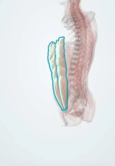
A group of conditions that arise in the muscles, sheath, nerves and embryological remnants of the anterior abdominal wall rather than in the organs behind it. The main ones are rectus sheath haematoma (bleeding into the rectus muscle from a torn epigastric artery, mostly in anticoagulated patients), diastasis recti (widening of the linea alba without a hernia defect), urachal and vitelline (omphalomesenteric) remnants at the umbilicus, desmoid tumour (a locally aggressive fibroblastic tumour), and anterior cutaneous nerve entrapment syndrome (ACNES), a common, overlooked cause of chronic localised abdominal pain.
Who gets it
ACNES is thought to explain a meaningful share of chronic abdominal pain, including in children and young women, and is often labelled functional pain. Rectus sheath haematoma is uncommon but rising with the use of anticoagulants and abdominal-wall injections of heparin; most patients are older women. Diastasis recti is very common after pregnancy and usually improves within the first months. Urachal remnants are found incidentally in a small fraction of children and adults on imaging. Desmoid tumours are rare, most often in women of reproductive age and in people with familial adenomatous polyposis.
Causes
- Rectus sheath haematoma: rupture of the inferior (or superior) epigastric artery or tearing of rectus fibres, triggered by anticoagulation, coughing, straining, vomiting, trauma, abdominal surgery or injections and trocar sites
- Diastasis recti: stretching and thinning of the linea alba by pregnancy (especially multiple or late pregnancies), obesity or ageing
- Urachal remnants: failure of the urachus (the channel between the fetal bladder and the umbilicus) to close: patent urachus, urachal sinus, urachal cyst or vesicourachal diverticulum
- Vitelline (omphalomesenteric) duct remnants: umbilical polyp, sinus, fibrous band or fistula to the ileum; Meckel diverticulum is the intestinal end of the same remnant
- Desmoid tumour: clonal fibroblastic proliferation driven by Wnt pathway mutations (CTNNB1 in sporadic cases, APC in familial adenomatous polyposis); pregnancy and surgical trauma are triggers
- ACNES: the anterior cutaneous branches of the lower intercostal nerves (T7 to T12) are compressed where they pass through small openings in the rectus sheath
Risk factors
- Anticoagulant or antiplatelet treatment, especially in older women, with cough or abdominal injections (haematoma)
- Pregnancy, multiple pregnancies, a large baby and obesity (diastasis, desmoid)
- Familial adenomatous polyposis and previous abdominal surgery (desmoid)
- Abdominal surgery, laparoscopic port sites and tight clothing or belts (nerve entrapment)
- Recurrent urinary tract infections or umbilical discharge in children (urachal remnant)
Pathophysiology
Haematoma: anticoagulation and a sudden contraction or cough → shear on the epigastric artery as it runs behind the rectus → bleeding inside the rectus sheath → below the arcuate line there is no posterior sheath, so blood can spread into the prevesical space and the pelvis → large haematomas cause anaemia, shock and abdominal compartment syndrome. Diastasis: stretching of the linea alba → widening between the two recti → a midline ridge on straining, but the fascia is intact, so nothing can strangulate. Nerve entrapment: the cutaneous nerve passes through a tight fibrous ring in the rectus sheath → compression and local ischaemia → pain at one point, made worse by tensing the muscle. Desmoid: local infiltrative growth of fibroblasts → a firm mass that invades muscle but never metastasises → recurs locally after incomplete excision.
Classification
- Rectus sheath haematoma by extent on CT: confined to the muscle; within the sheath and may cross towards the opposite side; spreading beyond the sheath into the prevesical space or peritoneum
- Urachal remnants: patent urachus (urine from the umbilicus), urachal sinus (blind tract opening at the umbilicus), urachal cyst (closed at both ends, may become infected), vesicourachal diverticulum (pouch at the bladder dome)
- Vitelline remnants: umbilical polyp, umbilical sinus, omphalomesenteric fistula (stool or mucus from the umbilicus), fibrous band (causes volvulus), Meckel diverticulum
- Desmoid tumour: abdominal wall, intra-abdominal (mesenteric, typical in familial adenomatous polyposis) and extra-abdominal
- Diastasis recti: graded by the width between the recti and by whether a true umbilical or epigastric hernia coexists
Symptoms and signs
- Rectus sheath haematoma: sudden severe pain and a tender, firm mass on one side of the abdomen, often after coughing; bruising around the umbilicus or flank may appear later
- ACNES: sharp or burning pain at one fingertip-sized point near the outer edge of the rectus, with tingling or altered skin sensation around it
- Diastasis recti: a soft midline ridge between the umbilicus and the xiphoid that bulges when sitting up; sometimes back pain and a sense of weak core
- Urachal remnant: clear or cloudy fluid, or an infected, red, tender swelling at or below the umbilicus
- Desmoid tumour: a firm, slowly growing, painless or mildly painful mass fixed to the muscle
Diagnosis
- Clinical
- Map the pain to a point and do Carnett sign. In the anticoagulated patient, a mass that stays palpable and does not cross the midline when the patient tenses the abdomen (Fothergill sign) suggests a rectus sheath haematoma. In ACNES, the pain is at one spot along the edge of the rectus, the skin around it is often hypersensitive to a pinch, and Carnett sign is positive. Diastasis is seen as a midline ridge when the patient lifts the head, with an intact fascial edge on palpation.
- Tests
- Haematoma: full blood count, coagulation tests and, in patients on warfarin, the INR; group and crossmatch. Infected umbilical remnant: urinalysis and culture of the discharge. Desmoid: core biopsy with beta-catenin staining, and colonoscopy if familial adenomatous polyposis is possible. ACNES: an injection of local anaesthetic at the tender point that abolishes the pain supports the diagnosis.
- Imaging
- Ultrasound is the first look for a wall mass, a haematoma, diastasis (measures the gap between the recti) or a urachal cyst. CT shows the size and spread of a rectus sheath haematoma, any active contrast blush, and rules out an intra-abdominal cause. MRI is the best study to map a desmoid tumour before deciding on treatment. In ACNES, imaging is normal and is done only to exclude other causes.
Differential diagnosis
- Incarcerated incisional, umbilical, epigastric or Spigelian hernia
- Intra-abdominal causes of acute pain (appendicitis, diverticulitis, ovarian torsion, cholecystitis)
- Pre-eruptive herpes zoster (band-like pain before the rash)
- Thoracic radiculopathy or diabetic truncal neuropathy
- Soft-tissue sarcoma, lipoma or metastasis in the abdominal wall
- Abdominal wall endometriosis in a caesarean scar (cyclic pain)
- Functional abdominal pain or irritable bowel syndrome (often wrongly given to ACNES)
Pain or a lump in the abdominal wall: which one?
| Condition | Typical patient | Bedside clue | Decisive test |
|---|---|---|---|
| Rectus sheath haematoma | Older woman on anticoagulants, after coughing | Tender one-sided mass that does not cross the midline and stays palpable on tensing | CT: haematoma in the rectus sheath, sometimes an active blush |
| Anterior cutaneous nerve entrapment | Young adult or child with months of pain | Fingertip-sized tender point at the rectus edge, Carnett positive | Pain relief after local anaesthetic at the point |
| Diastasis recti | After pregnancy | Soft midline ridge on sitting up, fascia intact | Ultrasound: widened gap between the recti, no hernia defect |
| Desmoid tumour | Young woman after pregnancy, or familial adenomatous polyposis | Firm, fixed, slowly growing mass | MRI and core biopsy |
| Incarcerated ventral hernia | Previous surgery or obesity | Tender irreducible bulge with a fascial defect | Ultrasound or CT: bowel or fat through the defect |
| Pre-eruptive herpes zoster | Older or immunosuppressed adult | Band-like burning pain in one dermatome | Rash appears within days |
Goal of treatment
Recognise that the pain or lump comes from the wall, treat the dangerous ones (expanding haematoma, infected remnant, growing tumour) actively, and spare the patient unnecessary abdominal surgery and endless investigations for the benign ones.
Treatment
- Acute
- Rectus sheath haematoma: most stop by themselves. Rest, analgesia, compression and ice; hold or reverse anticoagulation after weighing the reason it was prescribed (for example a mechanical valve or recent clot) with the specialist; transfuse if needed; treat the cough.
- Procedure
- Rectus sheath haematoma that keeps bleeding: angiographic embolisation of the epigastric artery; surgical evacuation and ligation only if embolisation fails or is unavailable, or if abdominal compartment syndrome develops.
- Procedure
- ACNES: injection of local anaesthetic, with or without a corticosteroid, at the tender point, which is both diagnostic and often therapeutic; if pain returns after repeated injections, anterior neurectomy (surgical division of the trapped nerve branch) relieves most patients.
- Rehabilitation
- Diastasis recti: reassurance that it is not a hernia and cannot strangulate; physiotherapy focused on deep abdominal and pelvic floor muscles. Surgical plication of the linea alba is an option for persistent symptoms or when a true hernia coexists.
- Surgical
- Urachal remnants: infected cysts are first drained and treated with antibiotics, then the whole remnant is excised, with a cuff of bladder in adults because of the small risk of urachal adenocarcinoma. Symptomatic vitelline remnants are excised, including any connection to the ileum.
- Follow-up
- Desmoid tumour: active surveillance with MRI is the first step for most, as many stop growing or shrink. Growing or symptomatic tumours are treated in a sarcoma centre with systemic therapy (NSAIDs, hormonal agents, tyrosine kinase inhibitors, gamma-secretase inhibitors), surgery or radiotherapy.
Complications
- Haemorrhagic shock, abdominal compartment syndrome and death from a large rectus sheath haematoma
- Thrombosis after anticoagulation is stopped
- Infection or abscess of a urachal or vitelline remnant; small bowel volvulus around a vitelline band
- Urachal adenocarcinoma in adults (rare)
- Local recurrence and invasion of neighbouring organs by desmoid tumours, especially mesenteric ones
- Unnecessary operations, imaging and chronic analgesic use when ACNES is missed
- Umbilical or epigastric hernia coexisting with diastasis recti
Common traps
- A positive Carnett sign proves the pain is functional: it means the source is in the abdominal wall, and that includes real disease such as a haematoma, a hernia or a nerve entrapment
- An anticoagulated patient with acute pain and a mass needs a laparotomy: a rectus sheath haematoma is managed without surgery in most cases; CT makes the diagnosis and embolisation stops ongoing bleeding
- Diastasis recti is a hernia that needs urgent repair: the fascia is intact and nothing can strangulate; physiotherapy is first, and surgery is elective and optional
- A desmoid is a benign tumour, so excise it straight away: it never metastasises but invades locally and recurs; many are now watched first because they may stop growing
- Chronic pain at one spot with normal tests is irritable bowel syndrome: a fingertip-sized point at the rectus edge with a positive Carnett sign is anterior cutaneous nerve entrapment, which a local anaesthetic injection can confirm and treat
Prognosis
Most rectus sheath haematomas resolve with conservative care, but large ones in older anticoagulated patients carry real mortality. ACNES has a good outlook once recognised: injections help many and neurectomy relieves most of the rest. Diastasis often improves after pregnancy and does not cause strangulation. Desmoid tumours do not metastasise; many stabilise, but mesenteric desmoids in familial adenomatous polyposis can be life-threatening.
Prevention
- Review anticoagulant intensity and treat cough in older patients; vary and correctly place abdominal-wall injection sites
- Do Carnett sign in every patient with chronic localised abdominal pain before ordering more imaging
- Excise symptomatic urachal and vitelline remnants completely
- Avoid unnecessary surgery on known desmoid tumours, as trauma can trigger regrowth
Test yourself
Think of the answer, then tap the question to see it.
A 74-year-old on an anticoagulant with a bad cough has sudden left lower abdominal pain and a tender mass still felt when she lifts her head. Likely diagnosis and next step?
Rectus sheath haematoma from a torn inferior epigastric artery. Check haemoglobin and coagulation, get a CT, hold or reverse anticoagulation with the specialist, and embolise if it keeps bleeding.
Why can a rectus sheath haematoma below the umbilicus spread into the pelvis?
Below the arcuate line the rectus has no posterior sheath, only transversalis fascia, so blood can track into the prevesical space and the pelvis.
A 24-year-old has had pain at one spot beside the right rectus for a year, with normal scans. The pain worsens when she tenses her abdomen. What is the diagnosis and how is it confirmed?
Anterior cutaneous nerve entrapment syndrome. Carnett sign is positive; an injection of local anaesthetic at the tender point that relieves the pain confirms it and often treats it.
A baby has clear fluid that smells of urine leaking from the umbilicus. What remnant is this?
A patent urachus, the persistent channel between the bladder dome and the umbilicus. Ultrasound confirms it, and it is excised.
Why are many desmoid tumours watched rather than removed at once?
They never metastasise, many stop growing or shrink on their own, surgery can trigger recurrence, and incomplete excision often fails.
Why it happens
Connections
Educational information, not medical advice. For any symptom, see a health professional.
Sources: Rectus Sheath Hematoma (StatPearls (NCBI Bookshelf), 2026); Diastasis Recti Rehabilitation (StatPearls (NCBI Bookshelf), 2023); Patent Urachus (StatPearls (NCBI Bookshelf), 2023); Desmoid Tumor (StatPearls (NCBI Bookshelf), 2024); Anatomy, Anterolateral Abdominal Wall Nerves (StatPearls (NCBI Bookshelf), 2023).
Spotted a mistake?We are always improving for the community. Tell us what is wrong and our team will review it.Report a mistake
Representative model, for educational purposes only.