
Pathological fracture and impending fracture (bone metastases and myeloma)
A bone weakened by cancer or another disease can break with an everyday load. Spotting the weak bone and fixing it before it snaps spares pain and surgery.
What it is
A fracture through bone already weakened by disease, occurring with normal activity or trivial trauma. In adults the commonest cause is metastatic carcinoma or multiple myeloma. An impending fracture is a lesion that has weakened a bone enough that it is likely to break under normal loads, and can be fixed before it does.
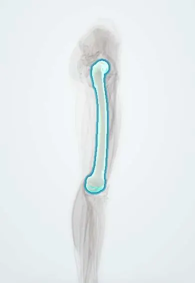
Who gets it
Bone is one of the commonest sites of metastasis, and most bone metastases come from breast, prostate, lung, kidney and thyroid cancer; multiple myeloma involves the skeleton in most patients. The spine is the commonest site, followed by the pelvis, proximal femur and proximal humerus. A sizeable minority of patients with bone metastases from breast cancer or myeloma will have a pathological fracture. In children, benign lesions such as bone cysts are the usual cause.
Causes
- Bone metastases: breast, prostate, lung, kidney and thyroid cancer most often
- Multiple myeloma and lymphoma of bone
- Primary bone tumours: osteosarcoma, Ewing sarcoma, chondrosarcoma; benign lesions such as unicameral bone cysts, enchondromas and non-ossifying fibromas (commonest in children)
- Metabolic bone disease: osteoporosis, osteomalacia, hyperparathyroidism, Paget disease, renal osteodystrophy
- Infection (osteomyelitis), radiation-damaged bone and osteogenesis imperfecta
- Long-term antiresorptive treatment: atypical femoral fractures
Risk factors
- Known cancer, especially breast, prostate, lung, kidney, thyroid or myeloma
- Lytic rather than blastic lesions
- Lesion in a weight-bearing bone, especially around the hip
- Pain on loading the limb (mechanical or functional pain)
- Large lesion destroying much of the cortex
- Previous radiotherapy to the bone
Pathophysiology
Tumour cells reach the marrow through the blood → they release factors that switch on osteoclasts (lytic lesions: kidney, thyroid, lung, myeloma) or osteoblasts (blastic lesions: prostate; breast often mixed) → osteoclast resorption hollows out the cortex, the part that carries load → a bone with a lytic hole loses strength far more than its size suggests, especially when the cortex is breached → pain appears first at rest from the tumour and then on loading as microfractures begin (mechanical pain) → a normal step, twist or lift breaks the bone. Healing is poor because tumour keeps destroying bone and radiotherapy slows repair, so the fixation must carry the load on its own for a long time.
Classification
- By cause: neoplastic (metastasis, myeloma, primary bone tumour) or non-neoplastic (metabolic, infection, radiation, genetic)
- Lesion type on X-ray: lytic, blastic or mixed
- Complete pathological fracture versus impending fracture
- Mirels score for long-bone metastases: site, pain, lesion type and size each scored 1–3 (total 4–12): site upper limb 1, lower limb 2, around the trochanters 3; pain mild 1, moderate 2, functional (with loading) 3; lesion blastic 1, mixed 2, lytic 3; size under one third of the bone's width 1, one to two thirds 2, over two thirds 3. A score of 9 or more favours prophylactic fixation; 7 or less, radiotherapy and observation; 8 is a judgement call
- Spine: the Spinal Instability Neoplastic Score (SINS) grades the stability of a vertebral metastasis
Symptoms and signs
- Bone pain that is worse at night and at rest, often present for weeks before the fracture
- Pain on weight-bearing or using the limb (functional pain), the warning of an impending fracture
- Sudden pain, deformity and loss of function after a minor event such as standing, turning or lifting
- Back pain with a vertebral collapse; weakness, numbness or bladder symptoms if the cord is compressed
- Symptoms of the underlying disease: weight loss, a breast lump, urinary symptoms, haematuria, cough, thirst and confusion from hypercalcaemia
Diagnosis
- Clinical
- Suspect it when the fracture is out of proportion to the trauma, when there was preceding pain, or in anyone with cancer. Ask about cancer history, weight loss and symptoms of a primary; examine the breasts, thyroid, prostate, lungs, abdomen and lymph nodes; check the neurology. In a patient with known metastases, ask about pain on loading the limb.
- Tests
- Full blood count, calcium, alkaline phosphatase, kidney function, serum and urine protein electrophoresis with free light chains (myeloma), PSA in men and other markers guided by the history. Biopsy of the lesion to confirm the diagnosis when the primary is unknown or the lesion is solitary, planned by the team that will operate.
- Imaging
- Plain X-rays of the whole bone, including the joints above and below, show lytic, blastic or mixed lesions and measure the cortical destruction. Staging with CT of chest, abdomen and pelvis and a bone scan; in myeloma a bone scan can be falsely normal, so use a skeletal survey, low-dose whole-body CT, MRI or PET-CT. MRI of the whole spine when vertebral involvement or cord compression is suspected.
Differential diagnosis
- Osteoporotic fragility fracture
- Ordinary traumatic fracture
- Primary bone sarcoma
- Stress fracture
- Atypical femoral fracture from long-term antiresorptive treatment
- Osteomyelitis
- Paget disease of bone
Bone broke too easily: what weakened it?
| Cause | Typical patient | X-ray look | Key test | Surgical rule |
|---|---|---|---|---|
| Metastatic carcinoma | Adult over 50, often with known breast, prostate, lung, kidney or thyroid cancer | Lytic, blastic or mixed lesions, often several | Staging CT and bone scan; biopsy if no known primary | Fix the whole bone, often prophylactically; radiotherapy afterwards |
| Multiple myeloma | Older adult with anaemia, kidney failure or high calcium | Multiple punched-out lytic lesions | Protein electrophoresis and free light chains; bone scan may be normal | Fix as for metastases; systemic treatment controls the disease |
| Primary bone sarcoma | Adolescent or young adult, or older adult with a solitary lesion | Aggressive solitary lesion with periosteal reaction | MRI of the whole bone and biopsy at a sarcoma centre | Never nail before biopsy; limb-salvage resection |
| Osteoporotic fragility fracture | Older adult, especially a woman after menopause | Thin cortices, no focal lesion | Bone density scan; exclude myeloma and metastases if in doubt | Standard fixation or arthroplasty; treat the osteoporosis |
| Benign bone lesion | Child or adolescent | Well-defined lucent lesion, such as a bone cyst | X-ray often diagnostic; MRI if unclear | Often heals with immobilisation; curettage and grafting if needed |
Goal of treatment
Relieve pain, restore function and walking quickly, prevent a fracture when one is impending, make fixation durable for the patient's expected survival without relying on bone healing, and treat the underlying disease.
Treatment
- Acute
- Initial care: analgesia, splinting, correction of hypercalcaemia and dehydration, and urgent spine assessment if there are neurological signs.
- Surgical
- Fixation that protects the whole bone and allows immediate weight-bearing: long intramedullary nails for shaft and trochanteric lesions; cemented hip replacement or tumour prostheses for femoral head and neck lesions, because these rarely heal.
- Surgical
- Prophylactic fixation of an impending fracture (for example a Mirels score of 9 or more, or pain on loading with a large lytic lesion): easier, with less blood loss and faster recovery than fixing after the fracture.
- Procedure
- Preoperative embolisation of very vascular metastases (kidney, thyroid) to reduce bleeding; biopsy before any fixation when the lesion is solitary or the primary is unknown.
- Procedure
- Radiotherapy to painful lesions that do not need fixation, and to the whole operated bone afterwards to control the tumour and protect the implant.
- Medical
- Bone-targeted drugs (bisphosphonates such as zoledronic acid, or denosumab) to reduce skeletal events, with dental review before starting; systemic treatment of the cancer or myeloma; analgesia including opioids when needed.
- Rehabilitation
- Early mobilisation and physiotherapy, walking aids and fall prevention; plan around the patient's goals and expected survival with the oncology and palliative care teams.
Complications
- Fixation failure or implant breakage when the bone does not heal
- Tumour progression around the implant
- Heavy bleeding during surgery on vascular metastases
- Fat or tumour embolism during reaming or cementing
- Spinal cord compression from vertebral collapse
- Hypercalcaemia, venous thromboembolism and pressure sores from immobility
- Osteonecrosis of the jaw from bone-targeted drugs
Common traps
- A fracture is a fracture, so nail it straight away: a solitary lesion with no known primary may be a sarcoma; nailing spreads it along the canal and can cost the limb, so stage and biopsy first
- A normal bone scan rules out bone disease in myeloma: myeloma lesions are purely lytic and often cold on bone scan; use a skeletal survey, whole-body CT, MRI or PET-CT
- A short plate across the broken segment is enough: metastatic disease affects the whole bone and fractures may never heal, so the implant must protect the whole bone and carry load by itself
- Night pain is the key sign of an impending fracture: rest pain reflects the tumour; pain with loading is what warns that the bone is about to fail
- A pathological fracture in a cancer patient means the end of active treatment: many live for years; fixation restores walking and quality of life even with limited survival
Prognosis
Survival depends on the cancer type: patients with breast, prostate, thyroid cancer or myeloma may live for years, while lung cancer metastases carry a shorter outlook. Many pathological fractures never heal, so outcome depends on durable fixation. Prophylactic fixation of an impending fracture gives better function and fewer complications than fixation after the bone has broken.
Prevention
- Regular review of patients with bone metastases, asking about pain on loading
- X-ray painful areas early in patients with cancer
- Use the Mirels score (long bones) and SINS (spine) to decide on prophylactic fixation
- Bone-targeted drugs in patients with bone metastases or myeloma
- Treat osteoporosis after any fragility fracture
Test yourself
Think of the answer, then tap the question to see it.
A 64-year-old with breast cancer has a lytic lesion destroying over two thirds of the width of her peritrochanteric femur and pain when walking. Mirels score and plan?
Site 3, functional pain 3, lytic 3, size 3: score 12. Prophylactic fixation (for example a long cephalomedullary nail), then radiotherapy to the whole femur.
Why is a bone scan unreliable for staging multiple myeloma?
A bone scan detects osteoblastic activity. Myeloma lesions are purely lytic with little new bone formation, so they often do not take up tracer.
A 58-year-old with no cancer history breaks his humerus lifting a bag; X-ray shows a solitary lytic lesion. Why should he not be nailed straight away?
The lesion may be a primary bone sarcoma. He needs staging and a planned biopsy first; nailing would contaminate the whole canal and could rule out limb-salvage surgery.
Why are pathological femoral neck fractures usually treated with cemented arthroplasty rather than screws?
The tumour-weakened neck rarely heals, especially after radiotherapy; replacing it gives a stable hip that bears weight at once without relying on bone healing.
Why it happens
- Bone tumours at a glance
- Osteosarcoma
- Chordoma, adamantinoma and marrow tumours in bone
- Fractures and fracture healing
- Bone metastases
- Pathways of spread and the usual metastatic sites
In radiology
Connections
Educational information, not medical advice. For any symptom, see a health professional.
Sources: Bone Metastasis (StatPearls (NCBI Bookshelf), 2023); Plasma Cell Neoplasms (Including Multiple Myeloma) Treatment (PDQ®)–Health Professional Version (National Cancer Institute, 2025); Osteosarcoma and Undifferentiated Pleomorphic Sarcoma of Bone Treatment (PDQ®)–Health Professional Version (National Cancer Institute, 2025).
Spotted a mistake?We are always improving for the community. Tell us what is wrong and our team will review it.Report a mistake
Representative model, for educational purposes only.