
Bile duct injury after cholecystectomy
Removing the gallbladder is routine, but cutting the main bile duct by mistake can scar the liver for life. A clear view before cutting is the best prevention.
What it is
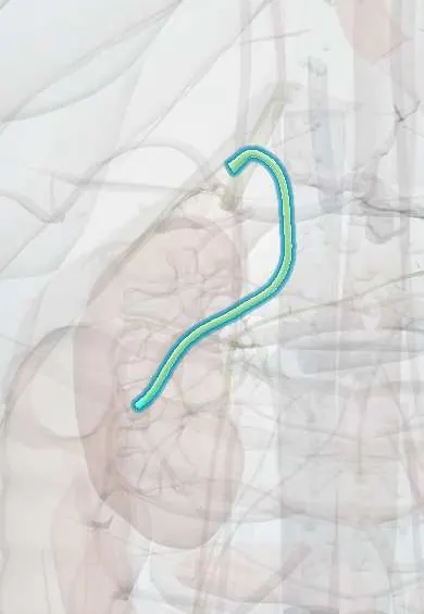
Damage to the extrahepatic bile ducts during gallbladder surgery, ranging from a leak from the cystic duct stump or a small duct in the liver bed to complete cutting, clipping or burning of the common bile or hepatic duct, sometimes with injury to the right hepatic artery. The page also covers the other causes of symptoms after cholecystectomy: retained bile duct stones and the postcholecystectomy syndrome.
Who gets it
Major bile duct injury complicates roughly 0.3–0.5% of laparoscopic cholecystectomies, about twice the rate of the open operation, and minor leaks are more frequent. Because cholecystectomy is one of the most common operations, the absolute number of injuries is significant. Gallstone disease is common in Colombia and in Latin America, and cholecystectomy is among the most frequent general surgery operations in the country. Only about a third of injuries are recognised during the operation.
Causes
- Misidentification: the common bile or hepatic duct mistaken for the cystic duct and clipped and cut
- Thermal injury from diathermy near the duct
- Clip slipping off the cystic duct stump or a leak from a small duct in the liver bed (duct of Luschka)
- Cutting an aberrant right hepatic duct that drains into the cystic duct or low into the common duct
- Tenting and tearing of the duct by excessive traction
- Ischaemic stricture after injury to the right hepatic artery or dissection that strips the duct's blood supply
Risk factors
- Acute or chronic inflammation: acute cholecystitis, a contracted fibrotic gallbladder, Mirizzi syndrome
- Anatomical variants of the ducts and arteries
- Bleeding that obscures the field and leads to blind clipping
- Obesity and difficult exposure
- Surgeon inexperience and failure to achieve the critical view of safety
- Emergency surgery and fatigue
Pathophysiology
Duct cut or clipped → if open, bile leaks into the peritoneum → biloma, bile peritonitis and sepsis over days; if clipped shut, bile cannot drain → obstructive jaundice and cholangitis → late healing of a partial or ischaemic injury → fibrous stricture → chronic obstruction → recurrent cholangitis, stones above the stricture, and over years secondary biliary cirrhosis and portal hypertension; associated right hepatic artery injury → ischaemia of the duct and part of the liver, making strictures more likely and repair less durable.
Classification
- Strasberg classification: A leak from the cystic duct stump or a duct of Luschka; B occlusion of an aberrant right hepatic duct; C transection of an aberrant right duct without ligation (leak); D lateral injury to the common duct; E circumferential injury to the main duct, subdivided E1–E5 by how close to the hepatic duct confluence it lies (the Bismuth levels)
- Minor injuries (Strasberg A, D) are usually managed endoscopically; major injuries (E) need surgical reconstruction
- Timing of recognition: during the operation, early after surgery (days) or late (stricture, months to years)
- Combined injury: bile duct plus vascular (right hepatic artery, portal vein)
- Postcholecystectomy syndrome: persistent or new upper abdominal symptoms after cholecystectomy, from biliary causes (retained or recurrent stones, cystic duct or gallbladder remnant, bile leak, stricture, sphincter of Oddi dysfunction) or non-biliary causes (peptic ulcer, reflux, pancreatitis, irritable bowel)
Symptoms and signs
- A patient who does not recover as expected after a routine laparoscopic cholecystectomy
- Abdominal pain, distension and loss of appetite in the first days
- Fever and fast pulse (bile peritonitis or biloma)
- Bile in the drain or leaking from a port site
- Jaundice, dark urine and pale stools (clipped or strictured duct)
- Months or years later: recurrent episodes of fever, rigors and jaundice (stricture with cholangitis)
Diagnosis
- Clinical
- During surgery: bile in the field, an unexpected extra duct, or a cholangiogram showing a missing or cut duct. After surgery: suspect injury in every patient who is not getting better within a few days, rather than attributing it to ordinary postoperative pain. Later: cholangitis or cholestasis in someone with a past cholecystectomy points to stricture or retained stones.
- Tests
- Liver function tests: rising bilirubin and alkaline phosphatase suggest obstruction, while a leak may show only a mild rise; full blood count and C-reactive protein for sepsis; bilirubin in drain fluid compared with serum to confirm a bile leak; blood cultures when febrile; amylase or lipase when pancreatitis from a retained stone is possible.
- Imaging
- Ultrasound or CT to find a collection or free fluid and dilated ducts. MRCP maps the biliary anatomy and the level of injury without instruments. ERCP shows leaks and lower duct injuries and can treat them (stent, sphincterotomy, stone removal); percutaneous transhepatic cholangiography shows the ducts above a complete transection and allows drainage. Intraoperative cholangiography defines anatomy and detects injury during surgery. CT angiography when an associated hepatic artery injury is suspected.
Differential diagnosis
- Retained or recurrent common bile duct stones
- Cystic duct stump or gallbladder remnant with stones
- Postoperative collection or abscess without bile leak
- Acute pancreatitis after surgery or from a passed stone
- Peptic ulcer, reflux or irritable bowel causing persistent pain (non-biliary postcholecystectomy syndrome)
- Malignant or sclerosing stricture (cholangiocarcinoma, primary sclerosing cholangitis) mistaken for a late benign stricture
Trouble after cholecystectomy: which one?
| Problem | When it shows | Typical picture | Key test | First move |
|---|---|---|---|---|
| Minor bile leak (cystic stump, duct of Luschka) | Days 1–7 | Pain, fever, bile collection; bilirubin normal or slightly raised | Ultrasound or CT, then ERCP | Drain the collection, ERCP stent to lower duct pressure |
| Major duct injury (cut or clipped common duct) | During surgery or first days to weeks | Bile peritonitis or progressive jaundice | MRCP, ERCP or percutaneous cholangiography | Control sepsis and drain; refer to a hepatobiliary centre for repair |
| Retained common bile duct stone | Weeks to months | Biliary colic, jaundice, sometimes pancreatitis | Ultrasound, liver tests, MRCP | ERCP with stone extraction |
| Cholangitis from stricture or stone | Any time, often late | Fever, rigors, jaundice, right upper quadrant pain | Cultures, liver tests, MRCP | Antibiotics and urgent biliary drainage |
| Non-biliary pain | Any time | Dyspepsia, reflux or bowel symptoms with normal liver tests | Endoscopy and clinical assessment | Treat the actual cause |
Goal of treatment
Prevent injury through safe technique; when it happens, recognise it early, control bile leak and sepsis, define the anatomy, and restore durable bile drainage in an experienced centre.
Treatment
- Procedure
- Prevention during surgery: achieve the critical view of safety (hepatocystic triangle cleared of fat and fibrous tissue, lower third of the gallbladder separated from the liver bed, only two structures entering the gallbladder), take a time-out before clipping, and use intraoperative cholangiography when anatomy is unclear.
- Surgical
- Bailout when the critical view cannot be reached: subtotal cholecystectomy, fundus-first dissection, cholecystostomy or conversion, accepting a lesser operation over the risk of a duct injury.
- Surgical
- Injury recognised during surgery: if a hepatobiliary surgeon is available, repair at once; if not, place drains and refer, without attempting a hasty repair, which often makes the injury worse.
- Acute
- Injury found after surgery: resuscitation, broad-spectrum antibiotics for sepsis, percutaneous drainage of collections and biliary decompression before any reconstruction.
- Procedure
- Endoscopic treatment for leaks and lateral injuries (Strasberg A and D): ERCP with sphincterotomy and stent to lower duct pressure so the leak heals; balloon dilation and stents for some strictures; stone extraction for retained stones.
- Surgical
- Major injury or complete stricture: tension-free Roux-en-Y hepaticojejunostomy with healthy, well-vascularised duct, done early or after inflammation settles, in a specialised hepatobiliary centre.
- Follow-up
- Long-term follow-up with liver tests and imaging to detect anastomotic stricture, recurrent cholangitis and progression to secondary biliary cirrhosis.
Complications
- Bile peritonitis and sepsis
- Biloma and intra-abdominal abscess
- Biliary stricture with recurrent cholangitis
- Secondary biliary cirrhosis and portal hypertension
- Liver atrophy or necrosis with combined arterial injury
- Stricture of the hepaticojejunostomy
- Reduced quality of life and medicolegal claims
Common traps
- The duct heading to the gallbladder must be the cystic duct: under traction the common bile duct can look exactly like it; only the critical view of safety proves which structure is which
- Converting to open surgery is a failure: when the critical view cannot be reached, conversion or a subtotal cholecystectomy is the safe, correct operation
- Slow recovery after a laparoscopic cholecystectomy is normal postoperative pain: patients should improve within a day or two, and one who is getting worse has a leak, injury or retained stone until proven otherwise
- The surgeon who caused the injury should repair it straight away: unless a hepatobiliary surgeon is present, drain and refer; hasty repairs fail more often and make later reconstruction harder
- Pain after cholecystectomy always comes from the bile ducts: many cases of postcholecystectomy syndrome are reflux, ulcer or irritable bowel; normal liver tests and imaging point away from the ducts
Prognosis
Minor leaks usually heal with endoscopic treatment. Major injuries repaired by experienced hepatobiliary surgeons have good long-term results in most patients, but outcomes are worse after repairs attempted outside specialised centres, after repeated repairs, with high injuries close to the confluence and with associated arterial injury. Untreated or failed strictures can lead to cirrhosis and the need for liver transplantation.
Prevention
- Critical view of safety before clipping or cutting any structure
- Bailout strategies (subtotal cholecystectomy, conversion) when the view cannot be obtained
- Intraoperative cholangiography or other imaging when anatomy is uncertain
- Careful use of energy devices near the ducts
- Training, supervision and a culture of stopping to ask for help
Test yourself
Think of the answer, then tap the question to see it.
What three conditions define the critical view of safety?
The hepatocystic triangle is cleared of fat and fibrous tissue, the lower third of the gallbladder is separated from the liver bed, and only two structures (cystic duct and artery) are seen entering the gallbladder.
Three days after a laparoscopic cholecystectomy a woman has increasing pain, fever and distension with a near-normal bilirubin. What is the likely problem and the first steps?
A bile leak, often from the cystic duct stump. Image for a collection (ultrasound or CT), drain it, then ERCP with a stent to lower pressure in the duct so the leak heals.
Why does an injury to the right hepatic artery make a bile duct injury worse?
The upper bile duct depends on arterial blood; losing it causes ischaemia of the duct and part of the liver, so strictures are more likely and repairs less durable.
A surgeon discovers a transected common bile duct during surgery in a hospital without a hepatobiliary surgeon. What is the safest action?
Place drains, avoid a hasty repair and refer to a hepatobiliary centre; early repair by an inexperienced surgeon often fails and makes later reconstruction harder.
Months after cholecystectomy a man has biliary colic and mild jaundice. Which cause is most likely, and how is it treated?
A retained or recurrent common bile duct stone; it is confirmed with liver tests and ultrasound or MRCP and removed by ERCP.
Why it happens
Connections
Educational information, not medical advice. For any symptom, see a health professional.
Sources: Bile Duct Injury (StatPearls (NCBI Bookshelf), 2023); Laparoscopic Cholecystectomy (StatPearls (NCBI Bookshelf), 2025); Postcholecystectomy Syndrome (StatPearls (NCBI Bookshelf), 2023); Choledocholithiasis (StatPearls (NCBI Bookshelf), 2023).
Spotted a mistake?We are always improving for the community. Tell us what is wrong and our team will review it.Report a mistake
Representative model, for educational purposes only.