
Vascular trauma: neck and limb arterial injuries
A cut or crushed artery can bleed to death in minutes or silently starve a limb or the brain. Hard signs mean theatre now; soft signs mean a scan first.
What it is
Injury to an artery or major vein from penetrating (stab, gunshot, glass, iatrogenic) or blunt (fracture, dislocation, crush, stretch) trauma. The injury may be a complete cut, a partial tear, an intimal flap with thrombosis, a pseudoaneurysm or an arteriovenous fistula. The bedside sorts patients by hard and soft signs: hard signs go straight to surgery, soft signs and stable penetrating neck wounds go to CT angiography.
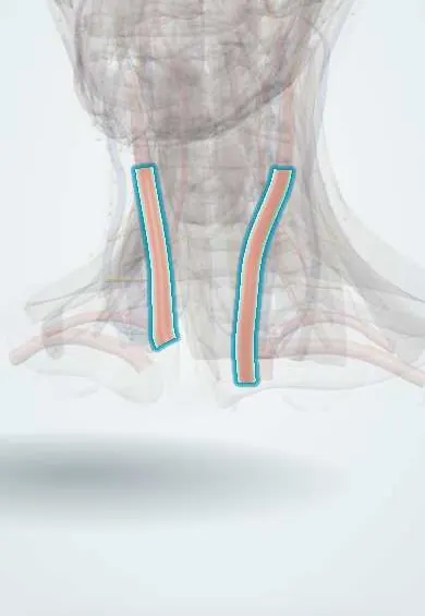
See it in 3D
Who gets it
Vascular injuries are a small fraction of all trauma but a large share of early haemorrhagic deaths. Most civilian injuries are penetrating and involve the limbs; the femoral, popliteal and brachial arteries are the most frequently injured in the extremities. In Colombia, firearm and stab wounds make penetrating neck and limb vascular trauma a frequent problem in urban emergency departments, mostly in young men.
Causes
- Gunshot wounds, including high-energy rifle and fragment injuries
- Stab wounds and cuts from glass or machetes
- Long-bone fractures and joint dislocations, especially knee dislocation (popliteal artery) and supracondylar humerus fracture in children (brachial artery)
- Crush and degloving injuries
- Blunt neck trauma with hyperextension or seatbelt injury (carotid and vertebral dissection)
- Iatrogenic: arterial punctures for catheterisation, central lines in the wrong vessel
Risk factors
- Penetrating wounds near the course of a major vessel
- Knee dislocation and high-energy fractures around the knee or elbow
- Combined arterial and venous injuries
- Delay to revascularisation beyond about 6 hours of ischaemia
- Associated bone, nerve and soft-tissue injury (mangled limb)
- Shock and coagulopathy from associated injuries
Pathophysiology
Vessel wall breached → free bleeding (shock) or contained bleeding (expanding haematoma, later pseudoaneurysm); artery and vein injured together → arteriovenous fistula with a thrill or bruit → or the intima tears and lifts as a flap → thrombosis and distal ischaemia → muscle and nerve tolerate only a few hours without flow → irreversible necrosis, and when flow returns, reperfusion swelling → compartment syndrome and release of potassium and myoglobin (rhabdomyolysis, kidney injury); in the neck, carotid or vertebral injury → stroke from occlusion or embolism, or airway compression by haematoma.
Classification
- Hard signs (operate): pulsatile bleeding, expanding or pulsatile haematoma, palpable thrill or audible bruit, absent distal pulses, signs of distal ischaemia (pain, pallor, paraesthesia, paralysis, coldness)
- Soft signs (investigate): history of significant bleeding at the scene, diminished but present pulse, small stable haematoma, wound close to a major vessel, adjacent nerve deficit, unexplained shock
- Neck zones (anatomical description): zone I from the clavicles and sternal notch to the cricoid cartilage, zone II from the cricoid to the angle of the mandible, zone III above the angle of the mandible to the skull base
- Penetrating neck injury means the wound breaches the platysma
- Injury types: transection, laceration, intimal flap or dissection, thrombosis, pseudoaneurysm, arteriovenous fistula, spasm (a diagnosis of exclusion)
- Mangled extremity: combined severe injury of at least three of bone, soft tissue, vessels and nerves; scores such as MESS describe severity but do not decide amputation alone
Symptoms and signs
- Bright red, pulsatile bleeding from a wound
- Swelling that grows or pulsates at the injury site
- A cold, pale limb with absent or weak pulses below the injury
- Numbness, tingling or weakness distal to the injury
- In the neck: hoarseness, stridor, difficulty swallowing, air bubbling from the wound, coughing or vomiting blood
- Focal neurological deficit (weakness, speech problems) after a neck injury
Diagnosis
- Clinical
- Primary survey first, with direct pressure or a tourniquet for limb bleeding. Then examine both limbs and compare pulses, colour, temperature, motor and sensory function. Hard signs mean surgery without delay. With soft signs, measure the ankle–brachial or arterial pressure index (injured limb systolic over uninjured arm): below 0.9 needs CT angiography. In the neck, check the platysma, airway, voice, swallowing and neurology; unstable patients or hard signs go to theatre, stable ones get imaging whatever the zone.
- Tests
- Haemoglobin, blood group and crossmatch, coagulation tests, blood gas with lactate; creatine kinase, potassium and renal function after prolonged ischaemia or crush (rhabdomyolysis); handheld Doppler signals and the pressure index at the bedside.
- Imaging
- CT angiography is the first imaging test for stable patients with soft signs and for stable penetrating neck injuries in any zone (the modern no-zone approach); it shows the arterial lesion, the wound track and the aerodigestive tract. Catheter angiography for unclear CT, artefact from metal fragments, or when endovascular treatment (stent graft, embolisation) is planned; on-table angiography in theatre. Duplex ultrasound for follow-up and some limb injuries. Contrast swallow or endoscopy when the track passes near the oesophagus.
Differential diagnosis
- Arterial spasm (diagnosed only after injury is excluded by imaging)
- Compartment syndrome without arterial injury
- Hypovolaemic shock causing weak pulses in all limbs
- Isolated nerve injury causing weakness and numbness with normal flow
- Laryngotracheal or oesophageal injury in penetrating neck trauma
- Pre-existing peripheral arterial disease with chronically absent pulses
Suspected arterial injury: findings and next step
| Situation | Typical findings | What it means | Next step |
|---|---|---|---|
| Hard signs (limb or neck) | Pulsatile bleeding, expanding haematoma, thrill or bruit, pulseless ischaemic limb | Arterial injury is near certain | Control bleeding, straight to theatre; on-table angiography if the site is unclear |
| Soft signs in a limb | Bleeding at the scene, weak pulse, small stable haematoma, wound near a vessel, nerve deficit | Injury possible, not proven | Pressure index; below 0.9, CT angiography |
| Stable penetrating neck wound through the platysma | No hard signs, normal airway and voice | Vessels, airway or oesophagus may be hit in any zone | CT angiography of the neck (no-zone approach), then targeted endoscopy or swallow study |
| Knee dislocation with normal-looking foot | Dislocation that may have reduced itself, pulses present | Popliteal intimal injury can thrombose hours later | Pressure index and serial pulse checks; CT angiography if abnormal |
Goal of treatment
Stop the bleeding, protect the airway in neck injuries, restore blood flow to the limb or brain within the ischaemic window, prevent compartment syndrome and save a limb only when it will be functional.
Treatment
- Acute
- Haemorrhage control: direct pressure, wound packing and a tourniquet placed high and tight on the limb; for junctional (groin, axilla, neck) bleeding, packing and pressure, sometimes a balloon catheter inserted into the wound; early balanced transfusion.
- Acute
- Neck injury with expanding haematoma or airway compromise: secure the airway early, before swelling makes intubation impossible, with a surgical airway ready.
- Surgical
- Open repair: proximal and distal control, removal of damaged wall and thrombus, then primary repair or an interposition graft (preferably the patient's own vein from the opposite leg); ligation is acceptable for some veins and non-essential arteries in damage control.
- Procedure
- Temporary intravascular shunt to restore flow quickly in damage control or before orthopaedic fixation of an unstable fracture, followed by definitive repair.
- Procedure
- Endovascular treatment (stent graft, embolisation) for selected injuries in difficult places such as zone I and zone III of the neck, the subclavian and axillary arteries.
- Surgical
- Fasciotomy of all compartments after prolonged ischaemia, combined arterial and venous injury or rising compartment pressure; primary amputation in a non-viable mangled limb after a multidisciplinary decision.
- Follow-up
- After repair: hourly distal pulses, Doppler signals, colour, temperature, motor and sensory checks, compartment assessment, urine output and colour; neurological checks after neck repair.
Complications
- Death from haemorrhage
- Limb loss and amputation
- Compartment syndrome and reperfusion injury
- Rhabdomyolysis and acute kidney injury
- Stroke after carotid or vertebral injury
- Airway obstruction from a neck haematoma
- Pseudoaneurysm or arteriovenous fistula found late
- Graft thrombosis or infection
Common traps
- A palpable distal pulse rules out an arterial injury: pulses can be present with an intimal flap, a partial tear or a pseudoaneurysm; measure the pressure index and image if it is low
- Every penetrating zone II neck wound needs mandatory exploration: stable patients without hard signs are now imaged with CT angiography whatever the zone, and only those with findings or instability are explored
- The cold pulseless limb is probably spasm: spasm is a diagnosis of exclusion; assume an arterial injury until imaging proves otherwise
- Fix the fracture first, then the artery: restore flow first, often with a temporary shunt, because the ischaemic clock is shorter than the time needed for fixation
- A tourniquet will cost the limb: a correctly applied tourniquet for a few hours rarely causes limb loss and saves lives; ischaemia from uncontrolled bleeding or delay is the real threat
Prognosis
Survival depends on early bleeding control; limb outcome depends mainly on the time to restored flow and on associated nerve and soft-tissue injuries. Revascularisation within about 6 hours gives the best chance of a functional limb. Neck injuries treated with early airway control and imaging-guided selective surgery have low rates of missed injury.
Prevention
- Violence prevention and control of firearms
- Road safety and workplace safety measures
- Early tourniquet use and bleeding-control training for first responders and the public
- Routine pulse checks and pressure index after knee dislocation and supracondylar fractures
Test yourself
Think of the answer, then tap the question to see it.
Which findings are hard signs of arterial injury, and what do they mean for the next step?
Pulsatile bleeding, expanding or pulsatile haematoma, palpable thrill or audible bruit, absent distal pulses and distal ischaemia. They make injury near certain: go to theatre, not to the scanner.
A man has a stab wound in zone II of the neck through the platysma, a normal voice and airway and no haematoma. What is the next step and why?
CT angiography of the neck. In stable patients without hard signs, imaging finds injuries in any zone and avoids unnecessary explorations; findings guide surgery, endoscopy or observation.
After a knee dislocation the foot pulses are present. Why is the patient not yet cleared, and what is measured?
An intimal tear of the popliteal artery can keep pulses at first and thrombose hours later. Measure the arterial pressure index and repeat pulse checks; a low index needs CT angiography.
Why is a temporary shunt placed before fixing an unstable fracture with an arterial injury?
It restores flow within minutes, so the muscle and nerve ischaemic clock stops while the bone is fixed; the definitive vessel repair is done afterwards at the correct length.
Why is a fasciotomy often done after revascularising a limb that was ischaemic for many hours?
Restored flow into damaged muscle causes reperfusion swelling inside closed compartments, which raises pressure and cuts off flow again (compartment syndrome).
Connections
Educational information, not medical advice. For any symptom, see a health professional.
Sources: Vascular Extremity Trauma (StatPearls (NCBI Bookshelf), 2023); Neck Trauma (StatPearls (NCBI Bookshelf), 2023); Injuries and violence (World Health Organization, 2024).
Spotted a mistake?We are always improving for the community. Tell us what is wrong and our team will review it.Report a mistake
Representative model, for educational purposes only.