
Cerebral cavernous malformation (cavernoma)
A small mulberry-like cluster of leaky vessels in the brain. Many never cause trouble; some cause seizures or small bleeds, and a few need surgery.
Quick facts
- Key signs
- Focal or focal-to-bilateral seizures · Sudden headache
- Diagnosis
- MRI
Red flags ↓
No doses: the clinician sets them by the current guideline.
What it is
A cerebral cavernous malformation is a cluster of enlarged, thin-walled capillary channels (caverns) lined by a single layer of endothelium, with no brain tissue between them and no feeding artery. It can occur anywhere in the brain or spinal cord. It presents with seizures, small repeated haemorrhages causing focal deficits, or as an incidental MRI finding. It is invisible on catheter angiography because the flow is so slow. Most are sporadic and single; familial forms, caused by mutations in KRIT1 (CCM1), CCM2 or PDCD10 (CCM3), are inherited dominantly and produce many lesions.
Who gets it
Cavernous malformations are found in about 1 in 200 people on brain MRI, most without symptoms. They present most often between 20 and 50 years of age but can be found in children. About four in five lie above the tentorium and up to a fifth in the brainstem. Familial cases account for a substantial minority; a founder mutation in KRIT1 is common among Hispanic families of Mexican ancestry, so familial disease should be considered in Latin American patients with several lesions or relatives with seizures or brain bleeds. Cranial radiotherapy in childhood can induce new cavernomas years later.
Causes
- Sporadic: usually a single lesion, often next to a developmental venous anomaly
- Familial: autosomal dominant loss-of-function mutations in KRIT1 (CCM1), CCM2 or PDCD10 (CCM3), with multiple lesions that increase over time
- Radiation-induced: new lesions years after cranial radiotherapy, especially in childhood
- Mechanism: a 'second hit' in endothelial cells disrupts the junctions between them → leaky, dilated caverns
Risk factors
- Family history of cavernomas, seizures or brain haemorrhage at a young age
- Previous haemorrhage from the lesion (the strongest predictor of another bleed)
- Brainstem location (higher bleeding risk and more severe consequences)
- Familial form, especially CCM3 (earlier and more aggressive disease)
- Cranial radiotherapy in childhood
Pathophysiology
Loss of function of a CCM gene in endothelial cells → weak endothelial junctions and abnormal signalling → dilated, thin-walled caverns without smooth muscle or elastin and without intervening brain → slow, low-pressure flow with thrombosis in the caverns → repeated small leaks of red cells → haemosiderin deposits and gliosis around the lesion → iron irritates the cortex → focal seizures (most often with temporal or frontal cortical lesions). A larger bleed or thrombosis enlarges the lesion → mass effect → sudden focal deficit, headache, or cranial nerve and long-tract signs in the brainstem. Bleeds tend to cluster: after one haemorrhage the risk of another is highest over the next couple of years. Because flow is slow and there is no arterial feeder → normal catheter angiography (angiographically occult).
Classification
- By presentation: incidental, seizures (epilepsy), symptomatic haemorrhage, focal deficit without haemorrhage
- By location: supratentorial (lobar, deep: basal ganglia and thalamus), infratentorial (brainstem, cerebellum), spinal cord
- Sporadic (single, often with a developmental venous anomaly) versus familial (multiple, gene mutation)
- MRI appearance (Zabramski types): acute or subacute blood, popcorn with mixed core and haemosiderin rim, chronic dark lesion, or tiny dot lesions seen only on susceptibility-weighted imaging
Symptoms and signs
- Focal or focal-to-bilateral seizures
- Sudden headache with a new neurological deficit after a bleed
- Weakness, numbness, visual loss or speech difficulty depending on site
- Brainstem lesions: double vision, facial weakness or numbness, unsteadiness, difficulty swallowing
- Symptoms that come in episodes and partly recover
- No symptoms at all (incidental finding)
Diagnosis
- Clinical
- Ask about seizures, episodes of focal deficit and recovery, headache, previous radiotherapy and family history of seizures, brain bleeds or cavernomas. A full neurological examination, including cranial nerves for brainstem lesions. In familial disease, examine relatives and look for skin vascular lesions, which some families have.
- Tests
- Electroencephalography in patients with seizures to relate the lesion to the seizure focus. Genetic testing for KRIT1, CCM2 and PDCD10 in patients with multiple lesions or a family history, with genetic counselling. Neuropsychological assessment before epilepsy surgery.
- Imaging
- MRI is the test of choice: a well-defined lesion with a mixed-signal core (blood of different ages) and a dark rim of haemosiderin on T2, the classic popcorn or mulberry appearance, with blooming on gradient-echo or susceptibility-weighted images, which are the most sensitive and reveal small additional lesions. MRI also shows an associated developmental venous anomaly. CT may show a subtle hyperdense or calcified lesion or acute blood, but can be normal. Catheter angiography is usually normal and is used only to exclude an arteriovenous malformation when the diagnosis is doubtful.
Differential diagnosis
- Arteriovenous malformation (flow voids, early venous filling on angiography)
- Haemorrhagic metastasis (melanoma, kidney, thyroid, choriocarcinoma)
- Hypertensive or amyloid intracerebral haemorrhage
- Low-grade glioma or other brain tumour
- Calcified neurocysticercosis
- Multiple microbleeds from hypertension or cerebral amyloid angiopathy
- Demyelinating lesion in the brainstem
A small bleed or seizure lesion on brain imaging: cavernoma or something else?
| Lesion | MRI or CT clue | Angiography | Bleeding pattern | Typical course |
|---|---|---|---|---|
| Cavernous malformation | Popcorn core with a dark haemosiderin rim; blooming on SWI | Normal (slow flow) | Small, repeated, low-pressure oozes | Seizures or episodic deficits; often observed |
| Arteriovenous malformation | Tangle of flow voids, enlarged draining veins | Nidus with early venous filling | Larger, high-pressure haemorrhage | Bleeds can be fatal; treated by surgery, embolisation or radiosurgery |
| Developmental venous anomaly | Caput medusae: radiating veins draining into one large vein | Normal arteries, umbrella of veins in the venous phase | Rarely bleeds by itself; often next to a cavernoma | Leave it alone: it drains normal brain |
| Haemorrhagic metastasis | Blood with surrounding oedema and enhancement; often several lesions | Normal or tumour blush | Bleeds into the tumour | Known or hidden primary cancer; grows |
| Hypertensive haemorrhage | Haematoma in basal ganglia, thalamus, pons or cerebellum | Normal | Single larger bleed | Older patient with long-standing hypertension |
| Calcified neurocysticercosis | Small calcified dots, sometimes with surrounding oedema | Normal | Does not bleed | Seizures in endemic regions |
Goal of treatment
Control seizures, prevent disabling repeated haemorrhage when the lesion is accessible and the risk justifies surgery, avoid harm from unnecessary operations on silent or deep lesions, and screen and counsel families.
Treatment
- Follow-up
- Incidental, asymptomatic lesions and many deep lesions: observation with clinical review and MRI if new symptoms appear. Patients learn the warning signs of a bleed.
- Medical
- Seizures: antiseizure medicines chosen as for focal epilepsy. Observational data suggest that antiplatelet or anticoagulant drugs, when needed for another reason, do not clearly raise the bleeding risk; the decision is individual. Drugs targeting the disease pathway are under study.
- Surgical
- Microsurgical resection: for accessible lesions causing drug-resistant or troublesome seizures (removing the lesion and the surrounding haemosiderin-stained brain improves seizure control), for symptomatic haemorrhage in accessible areas, and for brainstem lesions that have bled more than once and reach the surface of the brainstem, done in experienced centres.
- Surgical
- During surgery the associated developmental venous anomaly is preserved, because removing it can cause a venous infarct of the brain it drains.
- Procedure
- Stereotactic radiosurgery is considered for deep, inaccessible lesions that have bled repeatedly when surgery is too risky; its benefit is debated and it is not used for familial multiple lesions as routine.
- Lifestyle
- Familial disease: genetic counselling, testing of at-risk relatives and MRI with susceptibility-weighted imaging for those with symptoms or a confirmed mutation. Pregnancy and vaginal delivery are generally not considered to increase risk, and should be planned with the neurology team.
Complications
- Recurrent haemorrhage with stepwise neurological decline
- Drug-resistant epilepsy
- Permanent deficits after brainstem bleeds (diplopia, weakness, swallowing problems)
- Hydrocephalus from lesions near the fourth ventricle or aqueduct
- Surgical complications: new deficits, especially in brainstem surgery
- Psychological and driving or work restrictions linked to seizures
Common traps
- A normal cerebral angiogram rules out a vascular malformation: cavernomas are angiographically occult because flow is so slow; MRI with SWI is the test
- A cavernoma bleeds like an AVM and is a neurosurgical emergency every time: its bleeds are usually small low-pressure oozes; many lesions are observed
- The vein next to a cavernoma should be removed with it: the developmental venous anomaly drains normal brain; removing it can cause a venous infarct
- Removing the cavernoma alone is enough to stop seizures: the iron-stained brain rim is what irritates the cortex; resecting the rim improves seizure control
- A single cavernoma in a young patient needs no family history: multiple lesions or relatives with seizures or bleeds suggest a familial form that needs genetic counselling
Prognosis
Most incidental cavernomas never bleed in a lifetime, with a low yearly risk. The risk is clearly higher after a first haemorrhage and in the brainstem, and it is greatest in the first years after a bleed, then falls. Haemorrhages are usually small and rarely fatal, unlike those from arteriovenous malformations, but repeated brainstem bleeds can be disabling. Complete resection of an accessible lesion cures it, and most patients with seizures become seizure-free or improve after lesion and rim removal.
Prevention
- No way to prevent sporadic lesions
- Genetic counselling and family screening in familial disease
- Avoiding unnecessary cranial irradiation in children, and MRI follow-up after it
- Prompt review of new neurological symptoms in known carriers
Test yourself
Think of the answer, then tap the question to see it.
A 28-year-old has a first focal seizure. MRI shows a temporal lesion with a mixed core and dark T2 rim, blooming on SWI; catheter angiography is normal. What is it, and why is the angiogram normal?
A cavernous malformation. Its caverns have very slow, low-pressure flow and no feeding artery, so contrast does not fill them in a way angiography can show.
Why can a brainstem cavernoma cause disability from a bleed that would be silent in the frontal lobe?
The brainstem packs cranial nerve nuclei and long tracts into a tiny space, so even a few millimetres of blood or swelling causes diplopia, weakness or swallowing problems.
During resection of a cavernoma the surgeon sees a large draining vein beside it. Remove it?
No. It is a developmental venous anomaly that drains normal brain; removing it risks a venous infarct. Only the cavernoma and its haemosiderin rim are removed.
A 35-year-old woman has five cavernomas on SWI and her father had seizures. What next step besides treating her?
Suspect familial disease: offer genetic counselling and testing for KRIT1, CCM2 and PDCD10, and screen at-risk relatives.
Why it happens
Connections
Educational information, not medical advice. For any symptom, see a health professional.
Sources: Cerebral Cavernous Malformations (StatPearls (NCBI Bookshelf), 2025); Cerebral cavernous malformation (MedlinePlus Genetics (U.S. National Library of Medicine), 2025).
Spotted a mistake?We are always improving for the community. Tell us what is wrong and our team will review it.Report a mistake
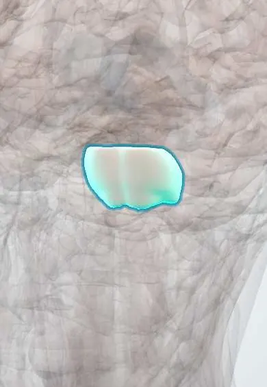
Representative model, for educational purposes only.