
Rhabdomyosarcoma
The commonest soft tissue cancer in children. It can start almost anywhere, so a growing lump, a bulging eye or a grape-like mass in a child needs a biopsy.
What it is
Rhabdomyosarcoma is a malignant tumour of cells that try, and fail, to become skeletal muscle. It is the commonest soft tissue sarcoma of childhood and adolescence, arising mostly in the head and neck, the genitourinary tract and the limbs, and it is treated with chemotherapy for every patient plus surgery and/or radiotherapy for the primary site.
Who gets it
Rhabdomyosarcoma makes up about 3% of childhood cancers and around half of childhood soft tissue sarcomas. Most cases occur before age 10, with a first peak in young children (embryonal type) and a second in adolescence (alveolar type, often in the limbs); it is rare in adults. In Colombia, childhood cancer is a notifiable event followed by the Instituto Nacional de Salud, and Law 1388 of 2010 gives children with suspected or confirmed cancer the right to prompt, comprehensive care; delays in reaching a paediatric oncology centre remain the main barrier to cure.
Causes
- Mostly sporadic, with no identifiable cause
- Alveolar type: a fusion of PAX3 or PAX7 with FOXO1 from a chromosomal translocation
- Embryonal type: no fusion; changes in RAS pathway genes and loss of heterozygosity on chromosome 11p15
- Cancer predisposition syndromes: Li–Fraumeni (TP53), neurofibromatosis type 1, Costello and Noonan (RAS pathway), Beckwith–Wiedemann, DICER1 syndrome
Risk factors
- Family history of early cancers (suggests Li–Fraumeni)
- Neurofibromatosis type 1
- RAS-pathway syndromes (Costello, Noonan)
- Beckwith–Wiedemann syndrome
- DICER1 syndrome (botryoid tumours of the cervix and bladder, pleuropulmonary blastoma in the family)
Pathophysiology
An immature mesenchymal cell starts the muscle programme (it makes desmin, myogenin and MyoD1) but cannot finish it → it keeps dividing as a primitive 'small round blue cell' → a mass grows in whatever tissue it sits in → its effects depend on the site: in the orbit → the eye is pushed forward early (found while small); next to the meninges (nasopharynx, sinuses, middle ear) → nasal blockage, ear discharge and cranial nerve palsies, and it can spread into the skull and the cerebrospinal fluid; in the bladder or prostate → urinary obstruction and blood in the urine; in the vagina of an infant → a grape-like mass protrudes (botryoid); beside the testis → a painless scrotal lump; in a limb → a growing lump, often with nodal spread. Cells spread early through lymphatics and blood → lymph nodes, lung, bone and bone marrow. Alveolar fusion-positive tumours → more metastases and a worse outcome.
Classification
- Histology: embryonal (commonest, younger children, head and neck and genitourinary; botryoid and spindle-cell variants have a good outlook), alveolar (adolescents, limbs and trunk, usually fusion-positive, worse), pleomorphic (adults) and spindle-cell/sclerosing
- Fusion status: PAX3/PAX7–FOXO1 fusion-positive behaves worse than fusion-negative, whatever it looks like
- Site: favourable (orbit, non-parameningeal head and neck, genitourinary other than bladder and prostate, biliary) versus unfavourable (parameningeal, bladder and prostate, limbs, trunk and others)
- Pretreatment stage: based on site, tumour size and invasion, nodes and distant metastases
- Clinical group: what remained after the first surgery, from complete removal to gross residual tumour or metastases
- Risk group: stage, group, histology or fusion status and age combined into low, intermediate and high risk, which set the intensity of treatment
Symptoms and signs
- Orbit: proptosis (bulging eye), swollen eyelid, displaced eye or limited eye movement, usually painless and growing over weeks
- Parameningeal (nasopharynx, sinuses, middle ear): nasal obstruction, nosebleeds, ear discharge or a polyp in the ear canal, facial swelling, cranial nerve palsies, headache
- Bladder or prostate: difficulty passing urine, retention, blood in the urine, constipation
- Vagina or cervix: a grape-like mass at the introitus or vaginal bleeding or discharge in an infant or young girl
- Paratesticular: a painless, firm lump above or beside the testis
- Limbs and trunk: a painless, growing lump; sometimes enlarged regional lymph nodes
- Metastatic disease: bone pain, cough, weight loss, pallor from marrow involvement
Diagnosis
- Clinical
- Suspect it in any child with a growing painless mass, unexplained proptosis, a mass in the ear, nose or vagina, or a painless scrotal lump. Examine the regional lymph nodes and the cranial nerves, and refer to a paediatric oncology centre before any surgery: the first operation should be a planned biopsy, not an attempt to 'shell out' the lump.
- Tests
- Biopsy (core needle or open) is essential: small round blue cells positive for desmin, myogenin and MyoD1 on immunohistochemistry, with molecular testing for the FOXO1 fusion. Staging tests: bilateral bone marrow aspirate and biopsy, cerebrospinal fluid cytology for parameningeal tumours, sampling of regional nodes for limb and paratesticular tumours (in older boys), blood count, kidney and liver function before chemotherapy, and genetic counselling when a predisposition syndrome is suspected.
- Imaging
- MRI (or CT) of the primary site to define size and invasion, including the skull base for parameningeal tumours. CT of the chest for lung metastases. PET-CT or bone scan to look for bone and nodal spread. Ultrasound is often the first study for a scrotal or abdominal mass.
Differential diagnosis
- Orbital cellulitis or chalazion (proptosis or eyelid swelling)
- Neuroblastoma metastatic to the orbit (bruising around the eyes)
- Other small round blue cell tumours: Ewing sarcoma, lymphoma, neuroblastoma, Wilms tumour
- Juvenile nasopharyngeal angiofibroma and nasal polyps
- Chronic otitis media with an aural polyp
- Hydrocele, hernia, testicular germ cell tumour
- Vaginal foreign body or benign polyp
- Benign soft tissue lumps (lipoma, haemangioma, fibromatosis)
A child with a lump: rhabdomyosarcoma or a look-alike?
| Condition | Typical age and site | Clue | First step |
|---|---|---|---|
| Rhabdomyosarcoma | Young children (head and neck, genitourinary) and adolescents (limbs) | Painless growing mass; desmin and myogenin positive | Planned biopsy at a paediatric oncology centre |
| Orbital cellulitis | Children of any age, after sinusitis | Fever, pain, red hot lid, rapid onset | Antibiotics and CT; reassess if it does not settle |
| Neuroblastoma | Under 5 years; adrenal, with orbital metastases | Bruising around the eyes, abdominal mass, raised urine catecholamines | Urine catecholamines, imaging, biopsy |
| Ewing sarcoma | Adolescents; bone of the pelvis and limbs, sometimes soft tissue | Bone pain, fever, onion-skin periosteal reaction; EWSR1 fusion | MRI and biopsy |
| Testicular germ cell tumour | Adolescents and young men; within the testis | Mass in the testis itself, raised tumour markers | Ultrasound and markers; inguinal approach |
Goal of treatment
Cure with the least lasting harm: confirm the diagnosis by biopsy, stage and assign a risk group, treat everyone with chemotherapy, and control the primary site with surgery and/or radiotherapy in a way that preserves the eye, the bladder and function wherever possible.
Treatment
- Medical
- Multi-agent chemotherapy for every patient, because microscopic spread is assumed; regimens are built on vincristine, dactinomycin and an alkylating agent (cyclophosphamide or ifosfamide), with more agents and maintenance for higher risk groups, given in a paediatric oncology centre within a protocol.
- Surgical
- Surgery for local control when the tumour can be removed completely without mutilation (for example many limb, trunk and paratesticular tumours, the latter through an inguinal approach). In the orbit, bladder, prostate and vagina, organ-sparing approaches after chemotherapy are preferred over radical removal.
- Procedure
- Radiotherapy for microscopic or gross residual disease, unresectable sites (orbit, parameningeal) and alveolar tumours, with conformal, proton or brachytherapy techniques chosen to spare growing tissues.
- Follow-up
- Supportive care during treatment: fever with neutropenia is an emergency, central line care, nutrition and fertility counselling. After treatment, imaging follow-up for relapse and long-term survivorship care for growth, fertility, heart, kidney and second cancers.
Complications
- Local recurrence or metastatic relapse (lungs, bone, marrow)
- Cranial nerve palsies and intracranial spread from parameningeal tumours
- Urinary obstruction and kidney damage from bladder or prostate tumours
- Febrile neutropenia and infections during chemotherapy
- Late effects: growth problems and facial asymmetry after radiation, cataract, infertility, bladder dysfunction, heart and kidney damage, second cancers
Common traps
- A soft tissue lump in a child can simply be excised to see what it is: an unplanned excision often leaves tumour behind and contaminates tissue planes; refer first and plan the biopsy
- Rhabdomyosarcoma only arises in skeletal muscle: it often arises where there is little muscle (bladder, prostate, vagina, middle ear, biliary tract), from immature mesenchymal cells
- A child with proptosis has orbital cellulitis until proven otherwise: painless, afebrile proptosis growing over weeks needs imaging and biopsy for rhabdomyosarcoma
- If the tumour is completely removed, chemotherapy is not needed: every patient receives chemotherapy because microscopic spread is assumed; surgery and radiation only control the primary site
- Alveolar and embryonal are just descriptive labels: fusion status (PAX3 or PAX7 with FOXO1) carries the prognosis and changes the risk group
Prognosis
About 70% of children with localised disease are cured, and low-risk tumours (such as orbital or paratesticular embryonal tumours) do better still. Fusion-positive alveolar tumours, parameningeal and limb sites, larger tumours, nodal spread and older age do worse. Metastatic disease and relapse carry a poor outlook, with fewer than one in three surviving.
Prevention
- No primary prevention is known
- Genetic counselling and surveillance for families with Li–Fraumeni, DICER1 and other predisposition syndromes
- Early recognition and prompt referral of children with unexplained masses
Test yourself
Think of the answer, then tap the question to see it.
A 2-year-old girl has a grape-like mass protruding from the vagina. What is the likely diagnosis and variant?
Embryonal rhabdomyosarcoma, botryoid variant, which grows beneath a mucosal surface (vagina, bladder, biliary tract) in young children and carries a relatively good prognosis.
A 6-year-old has painless proptosis growing over three weeks, no fever and no response to antibiotics. What next and why?
MRI of the orbit and a biopsy: this course does not fit infection and is typical of orbital rhabdomyosarcoma, which is treated with chemotherapy and radiotherapy, saving the eye.
Why does a child with a completely excised rhabdomyosarcoma still receive chemotherapy?
Rhabdomyosarcoma is assumed to have spread microscopically at diagnosis; before chemotherapy most patients relapsed after surgery alone.
A 15-year-old has a forearm mass with an alveolar pattern and a PAX3–FOXO1 fusion. How does this affect prognosis?
Fusion-positive alveolar tumour, a limb site and adolescence all predict a worse outcome, placing the patient in a higher risk group and calling for more intensive treatment.
Why is a paratesticular mass in a boy approached through the groin rather than the scrotum?
A scrotal incision opens a new lymphatic route and contaminates the scrotum; an inguinal approach removes the tumour with the spermatic cord, as for any testicular tumour.
Why it happens
- Rhabdomyosarcoma
- Other bladder tumours
- Diseases of the vagina
- Spermatic cord, paratesticular and tunica lesions
Connections
Educational information, not medical advice. For any symptom, see a health professional.
Sources: Rhabdomyosarcoma (StatPearls (NCBI Bookshelf), 2026); Childhood Rhabdomyosarcoma Treatment (PDQ®)–Health Professional Version (National Cancer Institute, 2025); Childhood Rhabdomyosarcoma Treatment (PDQ®)–Patient Version (National Cancer Institute, 2022).
Spotted a mistake?We are always improving for the community. Tell us what is wrong and our team will review it.Report a mistake
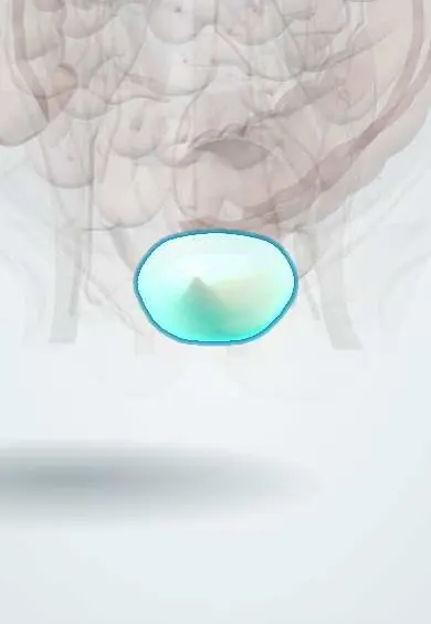
Representative model, for educational purposes only.