
Vertebrobasilar dolichoectasia
The arteries at the base of the brain grow too long, wide and twisted; they can cause strokes or press on the brainstem and facial nerves.
What it is
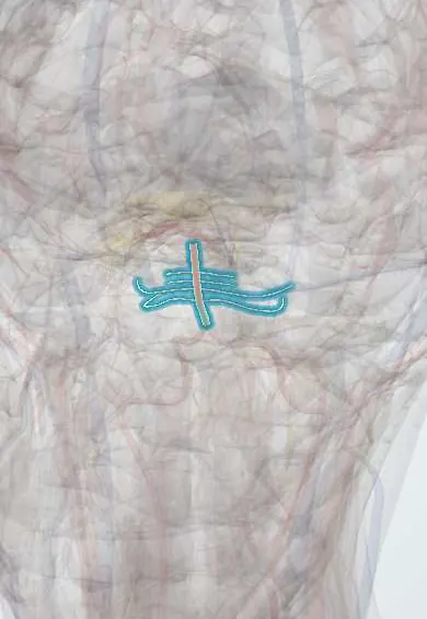
An arterial disease in which the vertebral and basilar arteries become abnormally long (dolicho-), wide (ectasia) and tortuous. It is often found by chance, but it can cause posterior circulation stroke, compression of the brainstem and cranial nerves, haemorrhage or hydrocephalus.
Who gets it
Uncommon, and estimates vary with the method: well under 1% in angiography and autopsy series, about 1.3% of people having routine MRI and MR angiography in one study, and a few per cent of people with posterior circulation infarcts. It is commonest in older men with hypertension.
Causes
- Degeneration of the arterial wall: fragmented internal elastic lamina and thin, atrophic smooth muscle
- Hypertension and atherosclerosis often coexist; whether they cause it or follow from it is debated
- Inherited and connective tissue conditions: Fabry disease, autosomal recessive polycystic kidney disease, Pompe disease, sickle cell disease, Marfan and Ehlers–Danlos syndromes
- Rarely infections of the vessel wall (syphilis, HIV, varicella-zoster virus)
- Growth of a fusiform aneurysm or an arterial dissection
Risk factors
- Older age
- Male sex
- Hypertension
- Atherosclerosis, diabetes and smoking (they raise the stroke risk once it is present)
- Dilated arteries elsewhere, such as an abdominal aortic aneurysm
Pathophysiology
The internal elastic lamina fragments and the media thins → the artery lengthens, widens and loops → flow slows and becomes to-and-fro, so mean flow falls → layered thrombus forms in the lumen and can embolise or block the mouths of perforating branches, while the stretched loop pulls and kinks those branches → infarcts in the brainstem, thalamus or posterior cerebral artery territory. Separately, the pulsating vessel acts as a mass → it compresses the pons, the medulla and the root entry zones of cranial nerves (trigeminal neuralgia, hemifacial spasm), or the floor of the third ventricle or the aqueduct (hydrocephalus). Repeated bleeding inside the vessel wall drives further growth, and a thin, fast-growing wall can rupture.
Classification
- By presentation: incidental (no symptoms), ischaemic, compressive, haemorrhagic, hydrocephalic
- By CT criteria (Smoker): elongation when the basilar artery runs outside the line of the clivus and dorsum sellae or its bifurcation lies above the suprasellar cistern; ectasia when its diameter is above about 4.5 mm
- By extent: basilar only, vertebral only or both; it may come with dilatation of anterior circulation arteries
Symptoms and signs
- Often none: it is found on imaging done for another reason
- Posterior circulation TIA or stroke: vertigo, double vision, unsteadiness, weakness or numbness
- Trigeminal neuralgia or hemifacial spasm from the pulsating artery pressing on the nerve root
- Other cranial nerve signs: double vision (abducens, oculomotor or trochlear nerve), tinnitus, hoarseness, difficulty swallowing
- Slowly progressive brainstem signs: weakness, unsteadiness, choking on liquids
Diagnosis
- Clinical
- Suspected when a posterior circulation stroke, a cranial neuralgia or slowly progressive brainstem signs occur, especially in an older man with hypertension; often an incidental finding. The diagnosis is made on imaging, since there are no universally agreed measurements.
- Tests
- No blood test makes the diagnosis. Vascular risk factors are checked (blood pressure, glucose, lipids); in a young patient an underlying cause such as Fabry disease may be sought. Evoked potentials and the blink reflex can show brainstem compression before symptoms appear.
- Imaging
- CT and CT angiography show the length, width, calcification and any thrombus in the lumen. MRI with MR angiography best shows the relation to the brainstem, cranial nerves and ventricles, and shows brainstem oedema and infarcts. Catheter angiography (DSA) is the reference standard and is used when treatment through the vessel is planned.
Differential diagnosis
- Saccular aneurysm of the basilar tip or a fusiform aneurysm
- Atherosclerotic vertebrobasilar stenosis
- Posterior fossa tumour, such as vestibular schwannoma or meningioma
- Classic trigeminal neuralgia from a small looping artery
- Hydrocephalus of other causes
A large basilar artery, or something else?
| Condition | Shape of the artery | Usual presentation | Key imaging clue |
|---|---|---|---|
| Vertebrobasilar dolichoectasia | Long, wide and tortuous along its whole course | Posterior stroke, cranial nerve compression, rarely bleeding | Wide basilar artery with its tip high above the suprasellar cistern or displaced to one side |
| Saccular aneurysm of the basilar tip | Normal vessel with a berry-like pouch | Rupture with a thunderclap headache (subarachnoid haemorrhage) | Focal outpouching at a branch point on CT angiography or DSA |
| Atherosclerotic vertebrobasilar stenosis | Narrowed, not widened | Repeated TIAs or stroke | Focal narrowing on CT or MR angiography |
| Classic trigeminal neuralgia | Normal-sized small artery looping onto the nerve | Electric facial pain alone | MRI shows a small vessel at the root entry zone and a normal basilar artery |
Goal of treatment
No treatment straightens the artery, so care targets its complications: preventing stroke, relieving compression, avoiding bleeding and treating hydrocephalus.
Treatment
- Medical
- Strict blood pressure control and management of the other vascular risk factors (diabetes, lipids, not smoking).
- Medical
- Antiplatelet or anticoagulant treatment may lower the risk of another ischaemic stroke but raises the risk of bleeding; the choice is made patient by patient, and the evidence is limited.
- Medical
- Medicines for trigeminal neuralgia or hemifacial spasm as in the classic forms (antiseizure medicines for neuralgia, botulinum toxin injections for spasm).
- Surgical
- Microvascular decompression for trigeminal neuralgia or hemifacial spasm that does not respond to medicines, in selected patients; it is harder than in classic cases because the artery is large and stiff.
- Surgical
- Ventriculoperitoneal shunt or endoscopic third ventriculostomy for obstructive hydrocephalus.
- Procedure
- Reconstruction through the vessel (overlapping stents, flow diverters) is still being evaluated and carries a high risk of brainstem infarction; it is reserved for progressive disease in specialised centres.
- Follow-up
- Follow-up imaging to watch for growth, wall thickening, thrombus or bleeding.
Complications
- Recurrent ischaemic stroke (the commonest cause of death)
- Subarachnoid or intracerebral haemorrhage, often fatal
- Progressive brainstem compression
- Obstructive hydrocephalus
- Hypertension from compression of the left ventrolateral medulla (a proposed mechanism)
Common traps
- Dolichoectasia is just atherosclerosis of an old artery: atherosclerosis often coexists, but the core lesion is a degenerated wall with a broken internal elastic lamina, and it also occurs in children and in inherited diseases
- A wide artery is above all a bleeding risk: ischaemic stroke is the commonest complication and cause of death; haemorrhage is rarer but often fatal
- Antithrombotic treatment is the safe default: it may reduce ischaemic recurrence but increases bleeding from the abnormal wall, so it is weighed case by case
- Trigeminal neuralgia always means a small looping artery: in an older patient with vascular risk factors a large dolichoectatic basilar or vertebral artery can be the culprit, and MRI shows it
Prognosis
Variable. Many incidental cases stay silent, but symptomatic disease tends to progress: in one long follow-up series median survival was under eight years, with ischaemic stroke the main cause of death and haemorrhage the most lethal event. Rapid growth and a thin wall predict bleeding.
Prevention
- Good blood pressure control
- Treating vascular risk factors and not smoking
- Follow-up imaging of known dolichoectasia to detect growth early
Test yourself
Think of the answer, then tap the question to see it.
What is the commonest serious complication of vertebrobasilar dolichoectasia?
Ischaemic stroke in the posterior circulation, from slow flow, thrombus in the lumen and kinked perforating branches; it is also the commonest cause of death.
A 70-year-old man with hypertension develops trigeminal neuralgia. MRI shows a wide, looping basilar artery touching the nerve root. What links the two?
The dolichoectatic artery pulses against the trigeminal root entry zone: the same mechanism as classic neuralgia, but with a much larger vessel.
How can vertebrobasilar dolichoectasia cause hydrocephalus?
The enlarged artery presses on the floor of the third ventricle or the aqueduct and blocks CSF flow; its pulsations may also hinder flow without a visible block.
Why is starting anticoagulation in dolichoectasia a difficult decision?
It may prevent another ischaemic stroke, but the abnormal wall can bleed and antithrombotic drugs raise that risk; benefit and harm are weighed for each patient.
Which imaging best shows how a dolichoectatic basilar artery relates to the brainstem and cranial nerves?
MRI with MR angiography. CT angiography shows shape and calcium well, and catheter angiography is kept for planning treatment.
Connections
Educational information, not medical advice. For any symptom, see a health professional.
Sources: Research Progress on Vertebrobasilar Dolichoectasia (International Journal of Medical Sciences (open access, PMC), 2014); Detection, cerebrovascular complications and risk factors associated with vertebrobasilar dolichoectasia: a scoping review (Frontiers in Neurology (open access, PMC), 2025).
Spotted a mistake?We are always improving for the community. Tell us what is wrong and our team will review it.Report a mistake
Representative model, for educational purposes only.