
Splenic lesions: cysts, abscess, infarct and splenic artery aneurysm
The spleen can develop cysts, pus, dead patches from blocked blood flow, or a ballooning artery that can burst, most dangerously in pregnancy.
What it is
Focal disease of the spleen and its artery, apart from trauma and simple enlargement. Splenic cysts are true (lined by epithelium, congenital), false (pseudocysts without a lining, after trauma, infarct or pancreatitis) or parasitic (hydatid). A splenic abscess is a collection of pus, usually seeded through the blood. A splenic infarct is death of part of the spleen when an artery branch is blocked by an embolus or a local clot. A splenic artery aneurysm is the commonest aneurysm of the abdominal organ arteries; most are small and silent, but rupture, above all in pregnancy, is life-threatening.
Who gets it
Splenic artery aneurysms make up about 60 percent of aneurysms of the abdominal organ arteries; they are commoner in women, especially after several pregnancies, and in portal hypertension and liver transplant candidates. Most ruptures in young women happen in pregnancy, usually in the third trimester. Splenic abscess is rare and mostly affects people with endocarditis, injection drug use, immunosuppression, diabetes or sickle cell disease, and, in the tropics, typhoid fever and malaria. Splenic infarcts follow endocarditis, atrial fibrillation, sickle cell disease and haematological cancers with big spleens. In Colombia, malaria, typhoid fever and sickle cell disease, more frequent in Pacific and Caribbean populations, are relevant causes.
Causes
- Cysts: congenital epithelial (true) cysts; pseudocysts after trauma, infarct or pancreatitis; hydatid cysts
- Abscess: bacteraemia from infective endocarditis or injection drug use, typhoid and other Salmonella; infection of an infarct or haematoma; spread from the pancreas, colon or stomach; fungi in neutropenia
- Infarct: emboli from the heart (atrial fibrillation, endocarditis, left ventricular clot); local clotting in sickle cell disease, myeloproliferative neoplasms, leukaemia and lymphoma with a large spleen; clotting disorders; twisting of a wandering spleen
- Splenic artery aneurysm: weakening of the artery wall with high flow, pregnancy, portal hypertension or fibromuscular dysplasia; false aneurysm (pseudoaneurysm) after pancreatitis or trauma
Risk factors
- Pregnancy and several previous pregnancies (aneurysm)
- Portal hypertension and cirrhosis
- Immunosuppression, HIV infection, diabetes and injection drug use (abscess)
- Atrial fibrillation, infective endocarditis and sickle cell disease (infarct)
- Chronic pancreatitis (pseudocyst, pseudoaneurysm)
Pathophysiology
Infarct: an embolus or local clot blocks an end-artery branch → the territory it supplies dies → a wedge-shaped infarct with its base on the capsule → left upper quadrant pain, sometimes pain at the left shoulder tip or on breathing; the infarct heals to a scar, liquefies into a pseudocyst, becomes infected (abscess) or rarely bleeds under the capsule and ruptures. Abscess: bacteria in the blood lodge in infarcted or damaged spleen → pus collects → fever, left upper quadrant pain and a left pleural effusion → rupture causes peritonitis. Aneurysm: the artery wall weakens (pregnancy hormones soften the middle layer, while blood volume, cardiac output and splenic flow rise; portal hypertension raises flow too) → the artery dilates → wall tension rises with diameter → rupture, sometimes first contained in the lesser sac and then free into the peritoneum ("double rupture") → haemorrhagic shock; in pregnancy it threatens both mother and fetus.
Classification
- Splenic cysts: primary (true epithelial, or parasitic) vs secondary (pseudocyst without lining)
- Splenic abscess: single vs multiple (multiple, small ones suggest fungi or immunosuppression)
- Infarct: segmental vs global (whole spleen)
- Splenic artery: true aneurysm vs pseudoaneurysm (after pancreatitis or trauma, higher risk of rupture)
- Other focal lesions to tell apart: haemangioma, hamartoma, lymphangioma, lymphoma and metastases
Symptoms and signs
- Often none: cysts and small aneurysms are usually found on imaging
- Left upper quadrant pain or fullness and early satiety with large cysts
- Pain at the tip of the left shoulder (Kehr sign) from irritation of the diaphragm
- Fever, rigors, left upper quadrant tenderness and a left pleural effusion in abscess
- Sudden left upper quadrant pain in infarct, sometimes worse on breathing
- Aneurysm rupture: sudden upper abdominal or left upper quadrant pain, then collapse and shock
Diagnosis
- Clinical
- Trace the lesion back to the blood: ask about heart disease and fever (emboli, endocarditis), sickle cell disease, blood cancers, pancreatitis, previous trauma, travel or rural exposure, immunosuppression and pregnancy.
- Tests
- Full blood count and blood film, inflammatory markers and several sets of blood cultures in fever or suspected abscess; echocardiography for endocarditis or a cardiac clot after an infarct; ECG or rhythm monitoring for atrial fibrillation; haemoglobin electrophoresis and clotting-disorder tests when unexplained; hydatid serology; pregnancy test in women of childbearing age; blood group and crossmatch when bleeding.
- Imaging
- Ultrasound for cysts and abscesses, and first in pregnancy and in the unstable patient to look for free fluid. Contrast CT: an infarct is a wedge-shaped area that does not enhance, with its base on the capsule; an abscess is a low-density collection with a rim, sometimes with gas; a cyst is a sharply outlined water-density lesion, sometimes with a calcified wall. CT angiography shows the size, neck and branches of an aneurysm and often wall calcification. MRI without gadolinium in pregnancy. Angiography is mainly used to treat.
Differential diagnosis
- Splenic injury and subcapsular haematoma
- Lymphoma or metastasis in the spleen
- Pseudocyst or tumour of the pancreatic tail
- Left renal colic or pyelonephritis
- Left lower lobe pneumonia or pulmonary embolism with pleuritic pain
- Placental abruption or uterine rupture (in pregnancy)
A focal lesion in the spleen: which one?
| Lesion | Typical patient | CT clue | Treatment |
|---|---|---|---|
| Splenic cyst | Young adult (true cyst) or after trauma or pancreatitis (false cyst) | Sharply outlined water density, thin wall, may calcify | Watch small ones; spleen-preserving surgery for large or symptomatic ones |
| Splenic abscess | Endocarditis, injection drug use, immunosuppression, sickle cell disease | Low-density collection with a rim, sometimes gas | Antibiotics and percutaneous drainage; splenectomy if they fail |
| Splenic infarct | Atrial fibrillation, endocarditis, sickle cell disease, myeloproliferative neoplasm | Wedge-shaped non-enhancing area with its base on the capsule | Pain relief and treat the cause, e.g. anticoagulation for an embolic source |
| Splenic artery aneurysm | Women after several pregnancies, portal hypertension | Round dilatation of the splenic artery, often with a calcified rim | Coiling, stent graft or surgery if 2 cm or more, growing, symptomatic or in pregnancy |
| Splenic injury | Blunt trauma to the left lower chest or abdomen | Laceration, haematoma, contrast blush | Observation, embolisation or splenectomy according to stability |
Goal of treatment
Keep the spleen whenever possible, treat the source of emboli or infection, and repair a splenic artery aneurysm before it ruptures, especially in women who are or may become pregnant.
Treatment
- Conservative
- Small asymptomatic cysts and small stable aneurysms in people who will not become pregnant: observation with periodic imaging.
- Medical
- Infarct: pain relief and treatment of the cause, such as anticoagulation for atrial fibrillation or a cardiac clot, antibiotics for endocarditis and care of sickle cell disease. Abscess: intravenous antibiotics guided by cultures, antifungals for fungal abscesses. Hydatid cyst: albendazole alongside surgery.
- Procedure
- Abscess: image-guided percutaneous drainage of a single abscess with one cavity. Splenic artery aneurysm: endovascular coil embolisation or a covered stent, the usual first choice in a stable patient.
- Surgical
- Large or symptomatic cysts: partial splenectomy or deroofing to keep splenic tissue. Splenectomy for multiloculated or failed abscess, bleeding, or an aneurysm at the hilum that cannot be treated endovascularly; other aneurysms can be resected or ligated with the spleen preserved.
- Acute
- Ruptured aneurysm: resuscitation with a massive transfusion protocol and emergency endovascular control or laparotomy with ligation of the splenic artery; in pregnancy, a joint obstetric and surgical team, often with caesarean delivery.
- Follow-up
- After splenectomy or loss of most splenic function: vaccines against pneumococcus, meningococcus and Haemophilus influenzae type b, and teaching to seek urgent care for fever (see asplenia).
Complications
- Rupture with intra-abdominal bleeding and shock
- Abscess rupture with peritonitis or left subphrenic abscess
- Infarct turning into a pseudocyst, abscess or subcapsular bleed
- Infection of a cyst
- Death of mother and fetus from aneurysm rupture in pregnancy
- Overwhelming infection after splenectomy
- Left pleural effusion
Common traps
- A small splenic artery aneurysm can always be watched: in a woman who is or may become pregnant any size is treated, because rupture in pregnancy is often fatal for mother and fetus
- Shock with abdominal pain in late pregnancy is always obstetric: a ruptured splenic artery aneurysm can mimic placental abruption or uterine rupture; think of it when the uterus does not explain the bleeding
- A splenic infarct is a final diagnosis: it is a sign of something else; look for the source, such as atrial fibrillation, endocarditis, sickle cell disease or a myeloproliferative neoplasm
- Every splenic lesion means splenectomy: cysts, abscesses and aneurysms can often be treated by drainage, partial resection or endovascular repair, keeping the spleen and its protection against infection
- A splenic abscess is a local problem: it usually arrives through the blood; search for endocarditis and treat the source along with the abscess
Prognosis
Cysts and infarcts usually do well, and the outcome follows the underlying cause. Splenic abscess still carries significant mortality when diagnosed late or in immunosuppressed patients. Ruptured splenic artery aneurysm has high mortality, and in pregnancy the risk to both mother and fetus is very high, so repairing at-risk aneurysms before they burst is the key.
Prevention
- Anticoagulation for atrial fibrillation when indicated
- Screen for and repair splenic artery aneurysms in women planning pregnancy, and report incidental ones
- Prevent and treat endocarditis, typhoid fever and malaria early
- Vaccinate before planned splenectomy, or after an emergency one
Test yourself
Think of the answer, then tap the question to see it.
A woman at 34 weeks collapses with sudden left upper abdominal pain; ultrasound shows free fluid and a normal placenta. What must be considered?
Rupture of a splenic artery aneurysm: an obstetric and surgical emergency needing resuscitation, massive transfusion, and laparotomy with control of the splenic artery and usually caesarean delivery.
Why do splenic infarcts look like wedges with their base on the capsule?
The splenic artery branches are end arteries that fan out toward the capsule, so a blocked branch kills a cone of tissue whose tip points to the hilum and whose base lies at the surface.
A man with fever, a new heart murmur and left upper quadrant pain has a splenic collection on CT. What else must be done?
Look for infective endocarditis with several sets of blood cultures and echocardiography: splenic abscesses and infarcts are frequent complications of endocarditis.
Why does pregnancy raise the risk of a splenic artery aneurysm rupturing?
Hormonal changes weaken the middle layer of the artery while blood volume, cardiac output and splenic flow rise, so wall tension increases, most of all in the third trimester.
Why is a spleen-preserving operation preferred for a large splenic cyst?
Removing the whole spleen leaves a lifelong risk of overwhelming infection by encapsulated bacteria; partial splenectomy or deroofing removes the cyst and keeps protective tissue.
Why it happens
Connections
Educational information, not medical advice. For any symptom, see a health professional.
Sources: Splenic Abscess (StatPearls (NCBI Bookshelf), 2025); Splenic Artery Aneurysm (StatPearls (NCBI Bookshelf), 2023); Spleen Imaging (StatPearls (NCBI Bookshelf), 2022); Splenomegaly (StatPearls (NCBI Bookshelf), 2025).
Spotted a mistake?We are always improving for the community. Tell us what is wrong and our team will review it.Report a mistake
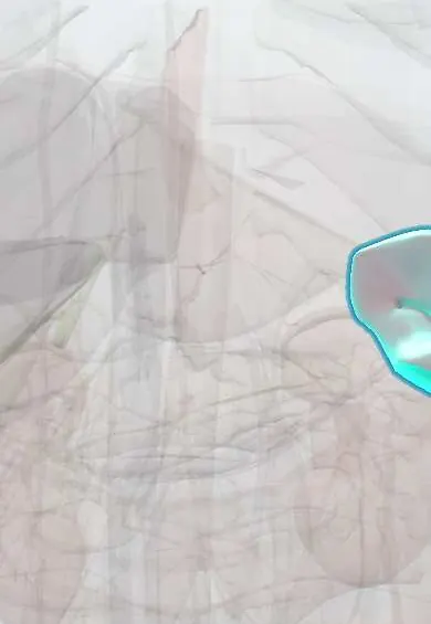
Representative model, for educational purposes only.