
Non-obstetric surgery and the acute abdomen in pregnancy
Pregnant women still get appendicitis, gallstones and bowel obstruction. Delaying surgery harms the baby more than a well-planned operation or a needed scan.
What it is
Any operation during pregnancy that is not for the pregnancy itself, most often for appendicitis, gallbladder disease, adnexal torsion, bowel obstruction, trauma or a breast mass. Care rests on a few principles: two patients but one circulation, imaging and anaesthesia chosen for safety but never withheld, laparoscopy adapted to the enlarged uterus, and close teamwork with obstetrics.
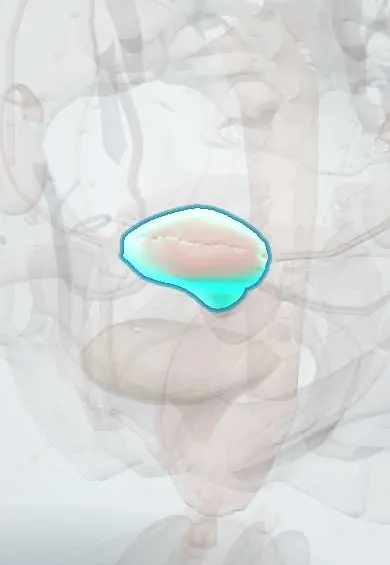
Who gets it
An estimated 0.2 to 2 per cent of pregnancies involve non-obstetric surgery. Appendicitis is the commonest non-obstetric surgical emergency in pregnancy, followed by biliary disease (biliary colic, cholecystitis, gallstone pancreatitis); adnexal torsion, bowel obstruction and trauma follow. In Colombia, where ultrasound is far more available than MRI outside large cities and many pregnant women live far from hospitals with obstetric and surgical teams, an ultrasound-first pathway and early referral are key; prenatal visits are a chance to explain which pains need urgent review.
Causes
- Acute appendicitis
- Gallstone disease: oestrogen raises cholesterol in bile and progesterone slows gallbladder emptying, so stones and sludge form
- Adnexal torsion, often of an enlarged ovary or cyst, most common in the first trimester
- Bowel obstruction from adhesions or volvulus, at times when the uterus changes size quickly
- Trauma (road traffic injuries, falls, interpersonal violence)
- A breast mass that may be pregnancy-associated breast cancer
Risk factors
- Previous abdominal surgery (adhesions)
- Known gallstones, obesity, rapid weight change
- Ovarian cysts or ovarian stimulation for fertility treatment (torsion)
- Delay in diagnosis because symptoms are blamed on the pregnancy
- Perforation of the appendix (sharply raises fetal loss and preterm birth)
- Family history of breast cancer or a breast lump that persists
Pathophysiology
The uterus grows out of the pelvis and displaces bowel, caecum and appendix upward and backward → pain may be felt higher and the peritoneum is less easily touched, so signs are softer → diagnosis is delayed → inflammation progresses (perforated appendicitis, gangrenous gallbladder, strangulated bowel) → maternal sepsis, hypotension and hypoxia reduce uterine blood flow, which has no autoregulation → fetal hypoxia, preterm labour and loss. Lying flat from about 20 weeks lets the uterus compress the inferior vena cava → less venous return → maternal hypotension under anaesthesia unless the uterus is tilted to the left. Pregnancy also brings lower oxygen reserve, slower gastric emptying and a more oedematous airway → faster desaturation and higher aspiration risk at induction.
Classification
- By urgency: emergency (never delayed for pregnancy), urgent or time-sensitive such as cancer surgery (best in the second trimester when possible), elective (deferred until after delivery)
- By trimester: first (organogenesis, highest spontaneous loss), second (preferred window when timing can be chosen), third (uterus in the way, preterm labour risk)
- By fetal viability: before viability, document the fetal heart rate before and after surgery; after viability, plan fetal monitoring and readiness for delivery with obstetrics
- Imaging by radiation: none (ultrasound, MRI without gadolinium) or ionising (X-ray, CT, fluoroscopy), used when the answer matters
Symptoms and signs
- Appendicitis: pain starting near the navel and moving to the right side, nausea, low fever; the pain is still most often in the right lower quadrant, though it may sit higher late in pregnancy
- Biliary colic and cholecystitis: right upper quadrant or epigastric pain after meals, fever and tenderness in cholecystitis
- Bowel obstruction: colicky pain, vomiting, distension, no flatus; vomiting must not be blamed on pregnancy
- Adnexal torsion: sudden one-sided lower abdominal pain with vomiting
- Breast mass: a firm, painless lump that persists, sometimes with skin change or a bloody nipple discharge
Diagnosis
- Clinical
- Confirm pregnancy and gestational age in every woman of reproductive age with abdominal pain or before surgery. Examine with her tilted to the left; tenderness may be higher and less marked. Separate surgical from obstetric causes (preterm labour, abruption, preeclampsia and HELLP, ectopic pregnancy) with early obstetric review and documentation of the fetal heart rate.
- Tests
- Interpret blood results against pregnancy norms: white cell count rises normally, alkaline phosphatase rises from the placenta and haemoglobin falls by dilution, so trends and the clinical picture matter more. Urinalysis, liver tests, lipase, blood pressure and platelets (to look for preeclampsia or HELLP), blood group and Rh status.
- Imaging
- Ultrasound first (appendix, gallbladder, ovaries, fetus and placenta). MRI without gadolinium when ultrasound is not diagnostic, especially for suspected appendicitis. CT is used when MRI is not available or the mother is unstable: the radiation from a single CT is well below the levels linked to fetal malformation, and iodinated contrast may be given when needed. For a breast mass: targeted ultrasound first, mammography with shielding if needed, and core needle biopsy.
Differential diagnosis
- Preterm labour
- Placental abruption
- Preeclampsia or HELLP syndrome (right upper quadrant pain)
- Ectopic pregnancy (first trimester)
- Acute pyelonephritis or kidney stones
- Degenerating uterine fibroid
- Round ligament pain
Abdominal pain in pregnancy: surgical or obstetric?
| Condition | Typical pain | Clue | First test |
|---|---|---|---|
| Appendicitis | Periumbilical, then right side; higher late in pregnancy | Fever, nausea, rising inflammatory markers | Ultrasound, then MRI without gadolinium |
| Acute cholecystitis | Right upper quadrant after meals, constant | Fever, Murphy sign, known gallstones | Ultrasound |
| Bowel obstruction | Colicky, central, with distension | Vomiting, no flatus, previous surgery | Ultrasound or MRI; plain films or CT if needed |
| Ovarian torsion | Sudden, one-sided, lower abdomen | Vomiting, known ovarian cyst, first trimester | Doppler ultrasound |
| Placental abruption | Constant pain, tense tender uterus | Vaginal bleeding (may be absent), fetal distress | Clinical and fetal monitoring; ultrasound may miss it |
| HELLP syndrome | Epigastric or right upper quadrant | High blood pressure, low platelets, raised liver enzymes | Blood pressure, platelets, liver tests |
Goal of treatment
Treat the mother's surgical disease as promptly and completely as outside pregnancy, while keeping her oxygenated, perfused and free of sepsis, protecting the fetus through the choice of technique and timing, and planning together with obstetrics, anaesthesia and neonatology.
Treatment
- Acute
- Resuscitation in left lateral tilt or with manual displacement of the uterus from about 20 weeks, oxygen, intravenous fluids and early antibiotics when infection is present; Rh-negative women receive anti-D immunoglobulin after abdominal trauma.
- Surgical
- Appendicitis: appendicectomy without delay, laparoscopic or open, in any trimester. Delay leading to perforation sharply raises fetal loss; antibiotics alone are not the standard in pregnancy.
- Surgical
- Biliary disease: laparoscopic cholecystectomy is safe in any trimester and is favoured for acute cholecystitis and recurrent symptoms, because conservative treatment often relapses; ERCP with minimal fluoroscopy for common bile duct stones or cholangitis.
- Surgical
- Laparoscopy adapted to pregnancy: open or left upper quadrant entry, ports placed according to fundal height, lower insufflation pressure, monitoring of maternal carbon dioxide, left tilt, and minimal handling of the uterus.
- Procedure
- Anaesthesia principles: no standard anaesthetic drug has been shown to cause malformations, and needed surgery must not be postponed for that fear. Regional anaesthesia where suitable; for general anaesthesia, preoxygenation, aspiration precautions and preparation for a difficult airway; avoid maternal hypotension, hypoxia and large swings in carbon dioxide.
- Follow-up
- Fetal heart rate documented before and after surgery; continuous fetal monitoring during surgery if the fetus is viable, the obstetric team agrees and delivery is possible. Watch for contractions afterwards; antenatal corticosteroids are given when preterm birth is likely, as decided by obstetrics. Thromboprophylaxis and early mobilisation.
- Medical
- Pain relief: paracetamol and short-term opioids; NSAIDs are avoided, particularly from about 20 weeks, because of effects on fetal kidneys and the ductus arteriosus.
- Surgical
- Breast mass: core needle biopsy without waiting for delivery; breast cancer surgery can be done in any trimester, chemotherapy is possible after the first trimester, and radiotherapy and some targeted and hormonal drugs are deferred until after delivery.
Complications
- Perforated appendicitis and peritonitis with maternal sepsis
- Miscarriage, preterm labour and preterm birth
- Fetal hypoxia or death from maternal hypotension or hypoxia
- Aspiration at induction of anaesthesia
- Venous thromboembolism
- Recurrent biliary attacks or gallstone pancreatitis with conservative management
- Delayed diagnosis and more advanced stage of pregnancy-associated breast cancer
Common traps
- In pregnancy the appendix pain moves to the right upper quadrant, so right lower quadrant pain is unlikely to be appendicitis: right lower quadrant pain remains the commonest presentation in every trimester; higher pain happens late in pregnancy
- A raised white cell count proves infection in pregnancy: it rises normally in pregnancy and labour; trends, fever and the clinical picture matter more
- CT is forbidden in pregnancy: ultrasound and MRI come first, but the radiation from a needed CT is far below the level linked to fetal harm, and a missed diagnosis is the greater danger
- Surgery should wait until after delivery if possible, even for cholecystitis: conservative treatment of acute cholecystitis often relapses with more risk to the pregnancy; laparoscopic cholecystectomy is safe in any trimester
- A breast lump in pregnancy is just hormonal change and can be checked after the baby is born: a persistent lump needs ultrasound and biopsy now; delay leads to more advanced pregnancy-associated breast cancer
Prognosis
With timely diagnosis and surgery, most mothers recover fully and most pregnancies continue to term. The fetal risk comes mainly from the disease and its delay (perforation, sepsis), not from imaging, anaesthesia or a careful operation.
Prevention
- Take abdominal pain in pregnancy seriously and examine the patient rather than attributing it to pregnancy
- Use an ultrasound-first imaging pathway and do not withhold MRI or CT when they will change management
- Plan elective surgery for after delivery and time-sensitive surgery for the second trimester when possible
- Teach pregnant women the warning signs that need urgent review
- Examine the breasts at the first prenatal visit and investigate any persistent lump
Test yourself
Think of the answer, then tap the question to see it.
A woman at 24 weeks has fever and right-sided abdominal pain; ultrasound cannot see the appendix. What next?
MRI without gadolinium if available; if not, or if she is unstable, CT. If appendicitis is confirmed, appendicectomy without delay, with left tilt, obstetric involvement and fetal heart rate monitoring.
Why should a pregnant woman beyond about 20 weeks be tilted to the left during surgery and resuscitation?
Lying flat lets the uterus compress the inferior vena cava, cutting venous return and cardiac output; maternal hypotension then reduces uterine blood flow, which the fetus depends on.
Why is laparoscopic cholecystectomy usually preferred over conservative care for acute cholecystitis in pregnancy?
Conservative treatment often relapses, with repeated admissions, pancreatitis and a higher risk of preterm labour; laparoscopic cholecystectomy is safe in any trimester.
Which drug class for pain is avoided especially after about 20 weeks of pregnancy, and why?
NSAIDs: they can reduce fetal kidney function (low amniotic fluid) and cause premature narrowing of the ductus arteriosus later in pregnancy.
At 18 weeks, a woman notices a firm breast lump that persists for three weeks. What is the right approach?
Do not wait for delivery: targeted breast ultrasound and core needle biopsy now, telling the pathologist she is pregnant; mammography with shielding if needed.
Connections
Educational information, not medical advice. For any symptom, see a health professional.
Sources: Appendicitis in Pregnancy (StatPearls (NCBI Bookshelf), 2026); Radiation Exposure In Pregnancy (StatPearls (NCBI Bookshelf), 2023); Physiology, Pregnancy (StatPearls (NCBI Bookshelf), 2023); New Palpable Breast Mass (StatPearls (NCBI Bookshelf), 2025).
Spotted a mistake?We are always improving for the community. Tell us what is wrong and our team will review it.Report a mistake
Representative model, for educational purposes only.