
Ureteral injury (iatrogenic and traumatic)
The ureter can be cut, tied or burned during pelvic surgery without anyone noticing. Days later, leaking or blocked urine causes pain, fever and swelling.
What it is
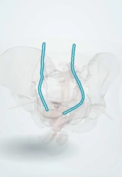
Damage to the ureter, the muscular tube carrying urine from the kidney to the bladder. Most injuries are iatrogenic, during gynaecological, colorectal, vascular or urological (ureteroscopic) surgery; a minority follow penetrating, rarely blunt, trauma. Injuries include ligation, crushing, partial or complete transection, thermal burns and devascularisation.
Who gets it
Ureteral injuries are rare compared with other urinary tract injuries but are the classic hidden complication of pelvic surgery. Hysterectomy is the commonest single cause, followed by colorectal and other pelvic operations; ureteroscopy is an increasing cause. Only a minority of iatrogenic injuries are recognised during the operation. External trauma injures the ureter in a small fraction of genitourinary trauma, mostly gunshot wounds, which in Colombia arrive together with other abdominal injuries.
Causes
- Gynaecological surgery: hysterectomy (abdominal, laparoscopic or vaginal), surgery for endometriosis, large fibroids or ovarian masses, caesarean section with extension
- Colorectal surgery: sigmoid colectomy and anterior or abdominoperineal resection of the rectum
- Urological surgery: ureteroscopy with perforation, stripping or avulsion
- Vascular, spinal and retroperitoneal surgery near the ureter
- Penetrating trauma: gunshot and, less often, stab wounds
- Blunt trauma with sudden deceleration, mainly avulsion at the ureteropelvic junction in children
Risk factors
- Distorted anatomy: endometriosis, large fibroids, malignancy, previous pelvic surgery or radiation, adhesions
- Heavy bleeding in the pelvis with blind clamping or suturing
- Use of energy devices close to the ureter
- Obesity and difficult exposure
- Congenital anomalies such as a duplex ureter
Pathophysiology
The ureter crosses three danger points: the pelvic brim, where it crosses the iliac vessels beside the ovarian vessels; the base of the broad ligament, where it runs under the uterine artery (water under the bridge) about 2 cm lateral to the cervix; and the vaginal cuff and bladder entry. A clamp, tie, cut or burn at one of these points → either urine leaks or flow is blocked. Leak → urine collects in the retroperitoneum or peritoneum (urinoma) → chemical irritation causes ileus and pain, infection causes fever and abscess, and the peritoneum reabsorbs urea and creatinine so serum creatinine rises even with a normal kidney. Blockage → pressure rises in the collecting system → hydronephrosis, flank pain and gradual loss of kidney function. Thermal or devascularisation injury → the wall dies days later → delayed leak or stricture. A tract to the vagina → ureterovaginal fistula.
Classification
- By timing of diagnosis: recognised during surgery (immediate) or delayed (days to weeks later)
- By mechanism: ligation or kinking, crush, partial or complete transection, thermal, devascularisation, avulsion
- By level: upper (to the pelvic brim), middle (over the iliac vessels) and lower (pelvic, to the bladder)
- AAST grades I–V: I haematoma or contusion without devascularisation; II tear of less than half the circumference; III tear of half or more; IV complete tear with less than 2 cm devascularised; V avulsion with more than 2 cm devascularised
Symptoms and signs
- Flank or loin pain on the affected side, or lower abdominal pain
- Fever, often in the first week after surgery
- Abdominal distension, nausea and prolonged ileus
- Increased watery output from a surgical drain or the wound
- Constant watery vaginal leak, days to weeks after hysterectomy, while the patient still voids
- Reduced urine output, or no urine at all if both ureters or a single kidney's ureter are tied
- Haematuria in some, but often absent
Diagnosis
- Clinical
- Suspect it after any pelvic or retroperitoneal operation that is followed by unexplained flank pain, fever, ileus, abdominal distension, watery drain output or vaginal leakage, and after penetrating wounds along the ureter's course. In the operating room, inspect the ureter's course when the field was difficult and look for leakage of intravenously given dye.
- Tests
- Serum creatinine and electrolytes (creatinine may rise from absorption of leaked urine); full blood count and C-reactive protein for infection; creatinine in drain or vaginal fluid, which is much higher than serum creatinine if the fluid is urine; urinalysis and urine culture. Intraoperative cystoscopy shows whether dye-stained urine jets from each ureteric orifice. A dye test with tampons in the vagina helps separate a ureterovaginal from a vesicovaginal fistula.
- Imaging
- CT urography with a delayed (excretory) phase is the main study: contrast extravasation, a urinoma, hydronephrosis or non-filling of the distal ureter. Retrograde pyelography during cystoscopy defines the level and allows a stent to be placed at once. Ultrasound shows hydronephrosis or a fluid collection and guides drainage. Antegrade pyelography through a nephrostomy maps the injury from above.
Differential diagnosis
- Bladder injury
- Vesicovaginal fistula
- Intra-abdominal abscess or haematoma
- Anastomotic leak after bowel surgery
- Postoperative ileus
- Acute kidney injury from hypotension or nephrotoxic drugs
- Ureteric stone or clot obstruction
- Lymphocele after pelvic lymph node dissection
Days after pelvic surgery: fever, pain or fluid. Where is it coming from?
| Problem | Typical clue | Key fluid or lab finding | Key imaging |
|---|---|---|---|
| Ureteral leak or obstruction | Flank pain, ileus, fever; constant vaginal leak if a fistula forms | Drain or vaginal fluid creatinine far above serum; serum creatinine may rise | CT urography delayed phase: extravasation, urinoma or hydronephrosis |
| Vesicovaginal fistula | Constant vaginal leak, often with little normal voiding | Dye instilled into the bladder stains a vaginal tampon | Cystoscopy and CT cystography show the bladder defect |
| Bladder injury | Haematuria, suprapubic pain, low urine output despite a catheter | Free fluid with urine-level creatinine | CT or conventional cystography with the bladder filled |
| Intra-abdominal abscess | Fever and pain from about the fifth day, rising inflammatory markers | Pus on drainage; drain creatinine similar to serum | CT: rim-enhancing collection |
| Anastomotic leak | Peritonitis, tachycardia and fever after bowel resection | Faeculent or turbid drain fluid | CT with rectal or oral contrast: leak at the anastomosis |
Goal of treatment
Recognise the injury as early as possible, divert or drain the urine to protect the kidney and control infection, and restore a tension-free, watertight, well-vascularised drainage from kidney to bladder.
Treatment
- Surgical
- Injury recognised during surgery: repair at once. A contusion or partial tear may need only a ureteral stent; a short gap in the upper or middle ureter is repaired end to end over a stent (ureteroureterostomy), with spatulated, tension-free ends.
- Surgical
- Lower ureteral injury: reimplantation into the bladder (ureteroneocystostomy), bridging longer gaps with a psoas hitch or a bladder flap (Boari flap). Long defects may need joining to the other ureter, an ileal ureter or kidney autotransplantation.
- Procedure
- Injury found late: attempt a retrograde stent; if it will not pass, place a percutaneous nephrostomy and drain the urinoma, then try an antegrade stent. Definitive repair is done early in a fit patient or once inflammation has settled.
- Acute
- Damage control in an unstable trauma patient: tie off or externalise the ureter and place a nephrostomy, leaving reconstruction for a later operation.
- Medical
- Antibiotics for an infected urinoma or obstructed infected kidney, and correction of fluids and electrolytes.
- Follow-up
- After repair: serum creatinine, drain output and its creatinine, stent removal on schedule, and later imaging (ultrasound or renography) to detect stricture and hydronephrosis.
Complications
- Urinoma and abscess
- Urinary sepsis
- Ureterovaginal or ureterocutaneous fistula
- Ureteral stricture and hydronephrosis
- Permanent loss of kidney function or loss of the kidney
- Acute kidney injury and anuria with bilateral injury
- Medicolegal claims, especially when recognition is delayed
Common traps
- No haematuria rules out a ureteral injury: haematuria is often absent; rely on imaging when the story fits
- A normal urine output means both ureters are intact: one injured ureter leaves the other kidney producing normal urine; only bilateral injury causes anuria
- A rising creatinine after surgery is always prerenal or drug-induced: urine leaking into the peritoneum is reabsorbed and raises serum creatinine
- Watery drain fluid after hysterectomy is just serous fluid or lymph: measure its creatinine; values far above serum mean urine
- A standard CT with contrast will show the leak: only a delayed excretory phase fills the ureter with contrast; request CT urography
Prognosis
Injuries recognised and repaired during the first operation do best, with high success rates. Delayed recognition means more operations, a higher risk of sepsis and stricture and a longer recovery, but most kidneys can still be saved with drainage and reconstruction.
Prevention
- Identify the ureter before clamping or dividing the uterine, ovarian and sigmoid vessels
- Know the three danger points: pelvic brim, under the uterine artery and the vaginal cuff
- Control pelvic bleeding with pressure and targeted ligation, not blind clamping
- Keep energy devices away from the ureter and avoid stripping its sheath
- Consider cystoscopy at the end of high-risk operations; preoperative stents help find the ureter but do not prevent injury
Test yourself
Think of the answer, then tap the question to see it.
Five days after a difficult hysterectomy, a woman has fever, ileus, left flank pain and a drain putting out watery fluid. What test on the drain fluid helps most?
Drain fluid creatinine: a value far higher than serum creatinine means the fluid is urine, pointing to a ureteral (or bladder) leak; confirm with CT urography.
Where does the ureter pass under the uterine artery, and why does it matter?
At the base of the broad ligament, about 2 cm lateral to the cervix. Clamping the uterine artery blindly at that point can tie or cut the ureter.
Why can serum creatinine rise after a unilateral ureteral leak despite a normal kidney on the other side?
Urine pooling in the peritoneum is reabsorbed across the peritoneal membrane, returning urea and creatinine to the blood.
Two weeks after hysterectomy, a woman has a constant watery vaginal leak but still voids normally. Dye in the bladder does not stain a vaginal tampon. Likely diagnosis?
Ureterovaginal fistula: urine from the injured ureter reaches the vagina directly, so bladder dye does not appear; a vesicovaginal fistula would stain the tampon.
Connections
Educational information, not medical advice. For any symptom, see a health professional.
Sources: Ureteral Injury (StatPearls (NCBI Bookshelf), 2023); Kidney and uro-trauma: WSES-AAST guidelines (World Journal of Emergency Surgery, 2019); Urotrauma Guideline - American Urological Association (American Urological Association, 2020); Hydronephrosis and Hydroureter (StatPearls (NCBI Bookshelf), 2023).
Spotted a mistake?We are always improving for the community. Tell us what is wrong and our team will review it.Report a mistake
Representative model, for educational purposes only.