
Sacrococcygeal teratoma
The most common newborn tumour grows from the tailbone. It is usually benign at birth, but it must come out early, coccyx and all, before it turns malignant.
What it is
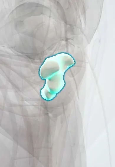
A sacrococcygeal teratoma is a germ cell tumour that arises at the tip of the coccyx and grows outward over the buttocks, inward into the pelvis, or both. It contains tissues from all three germ layers (skin, hair, neural tissue, gut, bone). It is the most common tumour of the newborn and is often found on prenatal ultrasound. At birth it is usually benign (mature or immature teratoma), but without complete removal, including the coccyx, it can recur or become malignant, most often as a yolk sac tumour.
Who gets it
About 1 in 35,000–40,000 live births; girls are affected 3–4 times more often than boys, but malignancy is relatively more common in boys. Most are now diagnosed on prenatal ultrasound in the second trimester. Tumours diagnosed before birth have a much higher perinatal mortality than those found after birth, mainly from fetal heart failure, prematurity and bleeding. Malignancy is uncommon in newborns but becomes frequent in tumours first found after the first months of life, often the hidden pelvic (type IV) ones.
Causes
- Thought to arise from pluripotent cells left near the coccyx (remnants of the primitive streak or misplaced primordial germ cells)
- Usually sporadic; a familial presacral form occurs in Currarino syndrome (presacral mass, anorectal malformation and sacral bone defect, MNX1 gene)
Risk factors
- Female sex (more frequent)
- Family history of Currarino syndrome (presacral teratoma)
- Late diagnosis and incomplete excision (risk of malignancy and recurrence)
Pathophysiology
Pluripotent cells at the coccyx → a mixed solid and cystic tumour → in the fetus, a large solid tumour with many vessels acts as an arteriovenous shunt → increased venous return and cardiac output → high-output cardiac failure, placental enlargement and hydrops → fetal death or premature delivery; the mother can develop mirror syndrome (oedema and pre-eclampsia-like signs mirroring the fetal hydrops). Bleeding into the tumour causes fetal anaemia; a large tumour can cause polyhydramnios, preterm labour and obstructed or traumatic delivery with rupture. Pelvic extension compresses the rectum, bladder, ureters and sacral nerves. Left in place (or if the coccyx is left), germ cells can give rise to a yolk sac tumour that secretes AFP.
Classification
- Altman type I: mostly external, minimal presacral part (the largest group)
- Altman type II: external with a significant pelvic part
- Altman type III: visible externally but mostly inside the pelvis and abdomen
- Altman type IV: entirely presacral, no external mass; diagnosed latest and most often malignant
- Histology: mature teratoma, immature teratoma (graded by immature neural tissue), malignant (mostly yolk sac tumour)
- Prenatal: predominantly cystic (better prognosis) versus solid and highly vascular (risk of heart failure and hydrops)
Symptoms and signs
- A mass at the bottom of the spine, between the coccyx and the anus, visible at birth; it may be huge, skin-covered or ulcerated
- Buttocks pushed apart, anus displaced forward
- Type IV: no visible mass; constipation, urinary retention, a lower abdominal mass or leg weakness in an infant or toddler
- Pallor and shock if the tumour bleeds
- Prenatal: an enlarging sacral mass on ultrasound, sometimes with polyhydramnios, a large placenta and fetal oedema
Diagnosis
- Clinical
- Most are suspected before birth. At birth, examine the mass, the skin over it and the anus; a digital rectal examination feels the pelvic part. Check the lower spine, legs, bladder and bowel function. In an older infant with constipation or urinary retention, a presacral mass felt on rectal examination is a type IV tumour until proven otherwise.
- Tests
- Serum AFP (very high in normal newborns, so the trend after excision matters more than one value) and β-hCG; blood count and coagulation in large or bleeding tumours. Histology of the whole specimen classifies mature, immature or malignant tissue.
- Imaging
- Prenatal ultrasound with Doppler shows a mixed solid and cystic sacral mass, its blood flow, and signs of heart failure, placental thickening or hydrops; fetal echocardiography follows cardiac output, and fetal MRI shows the pelvic extent. After birth, ultrasound and MRI (or CT) of the pelvis and abdomen define the intrapelvic part and the relation to the spinal canal, rectum and bladder; chest imaging if malignancy is suspected.
Differential diagnosis
- Myelomeningocele or lipomyelomeningocele
- Anterior sacral meningocele (Currarino syndrome)
- Lymphatic or vascular malformation
- Lipoma
- Rhabdomyosarcoma or neuroblastoma of the pelvis
- Rectal duplication cyst or abscess
Mass over the lower back of a newborn: tumour or neural tube defect?
| Lesion | Where exactly | Covering and content | Neurological signs | Key step |
|---|---|---|---|---|
| Sacrococcygeal teratoma | Below the sacrum, from the coccyx tip, often pushing the anus forward | Usually skin; mixed solid and cystic, may contain calcifications | Usually none at birth (pelvic types may compress nerves) | Early complete excision with the coccyx; AFP follow-up |
| Myelomeningocele | Over the lumbar or sacral spine, above the coccyx | Open neural placode leaking cerebrospinal fluid | Leg weakness, clubfeet, neurogenic bladder, hydrocephalus | Cover and close the defect; neurosurgery |
| Lipomyelomeningocele | Midline lumbosacral, above the gluteal cleft | Skin-covered soft fatty mass, often with a dimple, hair or haemangioma | May be normal at birth, deficits appear with growth (tethered cord) | Spinal MRI; neurosurgery |
| Lymphatic malformation | Buttock or perineum, not attached to the coccyx | Soft, cystic, transilluminating | None | Imaging; sclerotherapy or surgery |
Goal of treatment
Bring the fetus to a safe delivery, remove the whole tumour with the coccyx soon after birth, prevent bleeding, protect bowel, bladder and nerve function, and detect recurrence early.
Treatment
- Follow-up
- Prenatal: serial ultrasound and fetal echocardiography in a fetal medicine centre to follow tumour growth, blood flow, cardiac output, amniotic fluid and placenta; watch the mother for mirror syndrome.
- Procedure
- Fetal intervention in selected fetuses with heart failure or hydrops before they can be delivered: drainage of a large cyst or amniotic fluid, ablation of feeding vessels (laser or radiofrequency), or open fetal surgery to debulk the tumour, only in specialised centres.
- Acute
- Delivery planned in a centre with neonatal surgery; caesarean section for large tumours to avoid dystocia and rupture. After birth: protect the mass, keep the baby warm, secure venous access and blood for transfusion, and correct anaemia and clotting problems.
- Surgical
- Complete excision with the coccyx, early in the newborn period, through a posterior (chevron) buttock approach, combined with an abdominal approach for large pelvic parts (types III and IV); early control of the median sacral artery limits bleeding. The anal sphincter and pelvic floor are reconstructed.
- Medical
- Malignant tumours (yolk sac tumour) or malignant recurrence: platinum-based chemotherapy according to paediatric germ cell tumour protocols, followed by surgery for residual disease; outcomes are good with modern treatment.
- Follow-up
- Follow-up for at least 3 years with AFP, rectal examination and imaging at regular intervals, then long-term review of bowel, bladder and lower limb function.
Complications
- Fetal heart failure, hydrops and fetal death; maternal mirror syndrome
- Preterm birth and polyhydramnios
- Tumour rupture and massive bleeding during delivery or surgery
- Recurrence, more likely if the coccyx was left or the tumour was immature or spilled
- Malignant transformation (yolk sac tumour)
- Faecal incontinence, constipation and neurogenic bladder, more often with large pelvic tumours
- Wound infection and cosmetic problems of the buttocks
Common traps
- A benign teratoma at birth can be removed whenever convenient: the risk of malignancy rises steeply with age, so excision is done early in the newborn period
- Removing the visible tumour is enough: the tumour starts at the coccyx, and leaving the coccyx greatly increases recurrence
- A very high AFP in a newborn means the teratoma is malignant: AFP is physiologically high in newborns; what matters is the expected fall after excision and any later rise
- A mass at the base of the spine is always a neural tube defect: a teratoma lies below the sacrum at the coccyx tip, usually skin-covered and without leg weakness, while a myelomeningocele is higher, open and causes deficits
- The danger of a fetal teratoma is the tumour invading tissues: in the fetus the main threat is the tumour acting as a vascular shunt that drives high-output heart failure and hydrops
Prognosis
Babies with tumours found after birth and fully removed do very well. Prenatally diagnosed solid, vascular tumours with hydrops have a high perinatal mortality. Recurrence occurs in about 1 in 10 children, usually within 3 years, and may be malignant even when the first tumour was benign. Many survivors have some degree of bowel or bladder dysfunction.
Prevention
- No primary prevention
- Detailed second-trimester anatomy scan to find tumours early and plan delivery
- Excision soon after birth, always with the coccyx, to prevent malignant change and recurrence
- Rectal examination in infants with unexplained constipation or urinary retention (hidden type IV tumours)
- Structured AFP and imaging follow-up after surgery
Test yourself
Think of the answer, then tap the question to see it.
A fetus at 24 weeks has a large solid, very vascular sacral mass, a thick placenta and skin oedema. What is happening, and why?
High-output cardiac failure with hydrops: the tumour acts as a low-resistance arteriovenous shunt that steals cardiac output. It needs a fetal medicine centre; fetal intervention or early delivery may be considered.
Why must the coccyx be removed with a sacrococcygeal teratoma?
The tumour arises from pluripotent cells at the coccyx tip; leaving the coccyx leaves the source behind and greatly increases recurrence, which can be malignant.
A 14-month-old has worsening constipation and a firm presacral mass on rectal examination. What do you suspect and why is timing worrying?
An Altman type IV (entirely pelvic) sacrococcygeal teratoma, diagnosed late because nothing was visible. Tumours found after early infancy are often malignant (yolk sac tumour); check AFP and image urgently.
How is AFP used after excision of a sacrococcygeal teratoma in a newborn?
It should fall steadily toward normal for age; a rise after reaching that trend signals recurrence, often malignant yolk sac tumour, even with a normal examination.
Connections
Educational information, not medical advice. For any symptom, see a health professional.
Sources: Childhood Extracranial Germ Cell Tumors Treatment (PDQ®)–Health Professional Version (National Cancer Institute, 2025); Nonimmune Hydrops Fetalis (StatPearls (NCBI Bookshelf), 2025); Anesthetic Management of Ex Utero Intrapartum Treatment (EXIT) and Fetal Surgery (StatPearls (NCBI Bookshelf), 2025); Alpha-Fetoprotein Analysis (StatPearls (NCBI Bookshelf), 2025).
Spotted a mistake?We are always improving for the community. Tell us what is wrong and our team will review it.Report a mistake
Representative model, for educational purposes only.