
Spinal epidural and subdural hematoma
Bleeding inside the spinal canal that squeezes the spinal cord or nerves: sudden severe back pain, then weakness within hours. Surgery is urgent.
Quick facts
- Key signs
- Sudden · Radicular pain shooting into an arm · Rarely
- Diagnosis
- Emergency MRI of the suspect segment
Red flags ↓
No doses: the clinician sets them by the current guideline.
What it is
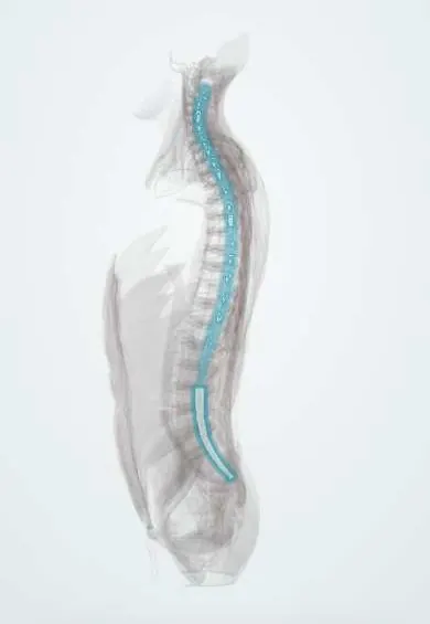
A collection of blood in the epidural space or, less often, the subdural space of the spinal canal that compresses the spinal cord, conus or cauda equina. It is rare and follows anticoagulant or thrombolytic treatment, spinal needles or surgery, or trauma, or arises with no cause; deficits can progress over minutes to hours.
Who gets it
Rare. Spontaneous spinal epidural hematoma has an estimated incidence of about 0.1 per 100,000 people per year, mostly in people in their sixties and seventies, with a slight male predominance. A symptomatic hematoma complicates about 0.3 to 1% of spine operations. Epidural collections are much more common than spinal subdural ones.
Causes
- Anticoagulant or thrombolytic treatment, and antiplatelet drugs
- Bleeding disorders and low platelet counts
- Lumbar puncture or neuraxial (spinal or epidural) anaesthesia in a patient with a bleeding tendency
- Spine surgery (postoperative hematoma)
- Spinal trauma, including vertebral fractures
- Vascular malformations and tumours
- Spontaneous, sometimes after straining, with no cause found
Risk factors
- Older age
- Anticoagulant, antiplatelet or thrombolytic drugs, especially in combination
- A spinal needle or catheter placed or removed while anticoagulated
- Multilevel spine surgery
- Coagulation defects or thrombocytopenia
- Pregnancy
Pathophysiology
Most hematomas come from the internal vertebral (epidural) venous plexus, whose veins have no valves and are exposed to pressure surges from straining; an arterial source is proposed for some cervical cases. Blood collects mostly behind or beside the dural sac over a few segments, with peaks at the cervicothoracic junction and the thoracolumbar region. Because the bony canal cannot expand, the clot compresses the spinal cord, its blood supply and the nerve roots: above the conus this causes a myelopathy, and below it a cauda equina syndrome. Prolonged compression and ischaemia turn a reversible deficit into a permanent one, which is why the time to decompression drives the outcome.
Classification
- By compartment: epidural (most) or subdural
- By cause: spontaneous, antithrombotic-related, procedure-related (lumbar puncture, neuraxial anaesthesia, surgery) or traumatic
- By level: cervical or thoracic (spinal cord compression) or lumbar (cauda equina compression)
- By deficit before surgery: incomplete or complete, the main guide to prognosis
Symptoms and signs
- Sudden, severe local neck or back pain, often with tenderness when the spine is tapped
- Radicular pain shooting into an arm, around the chest or down a leg
- Weakness of both legs, or of all four limbs in cervical bleeds, progressing over minutes to hours
- Numbness or tingling below a level on the trunk
- Urinary retention, bowel incontinence and saddle numbness when the cauda equina is compressed
- Rarely, weakness of one side of the body from a cervical clot, which can mimic a stroke
Diagnosis
- Clinical
- Suspect it in anyone with sudden back pain followed by spinal cord or cauda equina signs, particularly with anticoagulation, a bleeding disorder, a recent spinal procedure or trauma. Examine power, the sensory level, reflexes, perianal sensation and the bladder (post-void residual), and repeat the examination often because deficits can evolve quickly.
- Tests
- Coagulation studies (INR, activated partial thromboplastin time), platelet and blood counts, kidney function and blood group for surgery. These guide reversal and surgery but must never delay imaging.
- Imaging
- Emergency MRI of the suspect segment is the test of choice: it shows the clot, its compartment and its extent, and excludes abscess, tumour or a large disc. If MRI is not immediately available, CT myelography is used. Brain imaging done for a suspected stroke does not exclude a cervical spinal bleed.
Differential diagnosis
- Spinal epidural abscess
- Acute disc herniation with cauda equina syndrome
- Metastatic spinal cord compression
- Transverse myelitis
- Spinal cord infarction, including from aortic dissection
- Vertebral fracture
- Guillain-Barré syndrome
- Ischaemic stroke (when a cervical clot causes hemiparesis)
Back pain with a worsening deficit: which cause?
| Feature | Spinal epidural or subdural hematoma | Spinal epidural abscess | Acute disc herniation (cauda equina) | Metastatic cord compression |
|---|---|---|---|---|
| Typical setting | Anticoagulation, bleeding disorder, recent spinal needle or surgery, trauma | Bacteraemia, injection drug use, diabetes, recent spinal procedure | Lifting or straining, previous sciatica | Known cancer, often breast, lung, prostate or myeloma |
| Tempo | Minutes to hours | Days, often with fever | Hours to days | Days to weeks |
| Blood tests | Abnormal clotting or low platelets may be present | Raised inflammatory markers | Usually normal | Often normal; calcium may be raised |
| MRI | Epidural or subdural collection over a few segments, signal changing with clot age | Enhancing collection, often with discitis or osteomyelitis | Large disc fragment, usually low lumbar | Vertebral metastasis growing into the canal |
| First treatment | Reverse anticoagulation and decompress urgently | Antibiotics, usually with drainage | Urgent discectomy | Glucocorticoids, then radiotherapy or surgery |
Goal of treatment
Relieve compression before the deficit becomes permanent: stop and reverse antithrombotic drugs, and decompress urgently.
Treatment
- Acute
- Stop anticoagulant, antiplatelet and thrombolytic drugs and reverse them according to the agent: vitamin K with prothrombin complex concentrate for vitamin K antagonists (plasma if the concentrate is unavailable), idarucizumab or prothrombin complex concentrate for dabigatran, andexanet alfa or prothrombin complex concentrate for factor Xa inhibitors, and platelet transfusion for thrombocytopenia.
- Surgical
- Emergency decompressive laminectomy with evacuation of the hematoma. The shorter the time from symptom onset to surgery, the better the recovery, especially when the deficit is complete.
- Conservative
- Close observation without surgery only for small hematomas with mild or rapidly improving deficits, in a hospital with neurosurgery available, because some patients deteriorate later.
- Follow-up
- Frequent, structured neurological checks (power, sensation, bladder) after spine surgery and neuraxial anaesthesia and during observation; bladder catheter for retention; restarting thromboprophylaxis is weighed against rebleeding.
- Rehabilitation
- Spinal cord injury rehabilitation for residual deficits: physiotherapy, bladder and bowel programmes, pressure-area care and psychological support.
Complications
- Permanent paraplegia or tetraplegia
- Cauda equina syndrome with lasting bladder, bowel and sexual dysfunction
- Respiratory failure with high cervical compression
- Venous thromboembolism, pressure injuries and urinary infections during immobility
- Death, mainly with cervical hematomas in patients with cardiovascular disease or on anticoagulants
Common traps
- Back pain in an anticoagulated patient is muscular until proven otherwise: sudden severe back pain on anticoagulants, or after a spinal needle or surgery, needs a full neurological examination and urgent MRI
- Clotting must be corrected before imaging: reversal and MRI run in parallel, because every hour of compression worsens recovery
- Sudden hemiparesis always means a stroke: a cervical epidural hematoma can cause hemiparesis with neck pain, and giving thrombolysis or heparin can enlarge it
- A complete deficit makes surgery pointless: recovery is less likely than with an incomplete deficit but still happens with prompt decompression
Prognosis
Depends mainly on the severity of the deficit before surgery and the time to decompression. In spontaneous cases about 89% of patients with incomplete deficits recover fully within a year, compared with about 38% of those with complete deficits; mortality is around 6 to 8%, highest with cervical hematomas.
Prevention
- Respect the timing rules for stopping and restarting anticoagulants around lumbar puncture, neuraxial anaesthesia and catheter removal
- Check clotting and platelets before spinal procedures in patients at risk
- Structured neurological observation after spine surgery and during epidural analgesia, with a clear pathway to emergency MRI
- Avoid unnecessary combinations of antithrombotic drugs
Test yourself
Think of the answer, then tap the question to see it.
A woman on a factor Xa inhibitor gets sudden severe mid-back pain, and three hours later her legs are weak. What is the next step?
Emergency spinal MRI while the anticoagulant is stopped and reversed (andexanet alfa or prothrombin complex concentrate), with neurosurgery called for decompression. Reversal must not delay imaging.
Why do spinal epidural hematomas cause pain first and weakness later?
The growing clot first stretches the pain-sensitive dura and nerve roots; only as it enlarges in the rigid canal does it compress the cord or cauda equina, causing weakness, numbness and bladder failure.
Which factors best predict recovery after surgery for a spinal epidural hematoma?
The severity of the deficit before surgery (incomplete does far better than complete) and the time from onset to decompression; the earlier the operation, the better.
A patient with neck pain and sudden left hemiparesis has a normal head CT. Why should thrombolysis be withheld until the cervical spine is considered?
A cervical epidural hematoma can mimic a stroke with hemiparesis; head CT does not image the spinal canal, and thrombolysis or heparin can enlarge the clot.
When is non-surgical management of a spinal hematoma reasonable?
Only with small hematomas and mild or rapidly improving deficits, under close neurological observation in a hospital with neurosurgery, because some patients later worsen.
Connections
Educational information, not medical advice. For any symptom, see a health professional.
Sources: Spinal Subdural or Epidural Hematoma (MSD Manual Professional Edition, 2025); Spontaneous spinal epidural hematoma as a potentially important stroke mimic (Journal of Central Nervous System Disease (Europe PMC), 2014); Consensus statement by the Belgian Society of Neurosurgery and literature review on the diagnosis and management of postoperative spinal epidural hematoma (Brain and Spine (Europe PMC), 2024).
Spotted a mistake?We are always improving for the community. Tell us what is wrong and our team will review it.Report a mistake
Representative model, for educational purposes only.