
Retroperitoneal disorders: retroperitoneal fibrosis, haematoma and sarcoma
Behind the abdominal lining is a hidden space that can fill with scar, blood or a huge tumour before anyone notices. Kidneys, ureters and big vessels sit in it.
Quick facts
- Key signs
- Fibrosis · Fibrosis · Fibrosis
- Diagnosis
- Contrast CT
- First line
- Fibrosis
Red flags ↓
No doses: the clinician sets them by the current guideline.
What it is
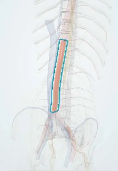
The retroperitoneum is the space behind the parietal peritoneum that holds the kidneys, ureters, adrenals, aorta, inferior vena cava, pancreas, parts of the duodenum and colon, and the psoas muscles. Three surgical problems are typical. Retroperitoneal fibrosis is inflammatory scar tissue that wraps the lower aorta and iliac vessels and traps the ureters. Retroperitoneal haematoma is bleeding into the space from trauma, anticoagulation, a leaking aneurysm, a kidney tumour or a high femoral puncture. Retroperitoneal sarcoma is a malignant soft-tissue tumour, most often liposarcoma or leiomyosarcoma, that grows very large before it is found.
Who gets it
Retroperitoneal fibrosis is rare, about one per 100 000 people a year, commonest in men aged 40–60; most cases are idiopathic and many are part of IgG4-related disease. Retroperitoneal haematoma is common after major blunt trauma, especially pelvic fractures, and in older patients on anticoagulants. Retroperitoneal sarcomas make up about 10–15% of soft-tissue sarcomas and are often 15 cm or more at diagnosis. In Colombia, road-traffic injuries (many from motorcycles) are a major cause of trauma-related retroperitoneal haematoma, and tuberculosis remains a cause of secondary fibrosis to exclude.
Causes
- Fibrosis, idiopathic: immune-mediated periaortitis, often IgG4-related disease
- Fibrosis, secondary: drugs (ergot derivatives such as methysergide, some dopamine agonists), inflammatory abdominal aortic aneurysm, malignancy (lymphoma, metastatic cancer), radiotherapy, infections such as tuberculosis, previous surgery
- Haematoma: blunt or penetrating trauma, pelvic fracture
- Haematoma: anticoagulant or antiplatelet therapy, coagulopathy, haemodialysis
- Haematoma: ruptured abdominal aortic or visceral artery aneurysm, renal angiomyolipoma or renal cell carcinoma (Wunderlich syndrome), femoral puncture above the inguinal ligament
- Sarcoma: liposarcoma (well-differentiated and dedifferentiated), leiomyosarcoma (often from the inferior vena cava), solitary fibrous tumour and others; previous radiotherapy and some hereditary syndromes in a few
Risk factors
- Male sex and age 40–60 (fibrosis)
- Smoking and asbestos exposure (fibrosis)
- Anticoagulation, especially when combined with antiplatelet drugs or kidney failure (haematoma)
- Femoral arterial access for catheterisation (haematoma)
- High-energy trauma, pelvic fracture (haematoma)
- Previous radiotherapy to the abdomen (sarcoma)
Pathophysiology
Fibrosis: immune inflammation around the infrarenal aorta → a plaque of fibrous tissue spreads laterally over the iliac vessels → it encases the ureters and pulls them toward the midline → urine flow is blocked → bilateral hydronephrosis and kidney failure; the same plaque compresses the inferior vena cava and gonadal veins → leg oedema, deep vein thrombosis, varicocele. Haematoma: bleeding into the loose retroperitoneal space → large volumes can collect before peritoneal signs appear (several litres) → hypovolaemia with few abdominal findings → blood tracks to the flanks (Grey Turner sign) and along the psoas → femoral nerve compression → thigh numbness and weak knee extension. Sarcoma: slow growth in a space with room to expand → silent until very large → displaces organs and vessels → fullness, early satiety, leg oedema → it recurs locally from the edges if not removed whole, and local recurrence causes most deaths.
Classification
- Retroperitoneal fibrosis: idiopathic (including IgG4-related) or secondary (drugs, aneurysm, cancer, radiation, infection, surgery)
- Traumatic haematoma by zone: zone I central (aorta, vena cava, pancreas, duodenum), zone II lateral (kidneys, colon), zone III pelvic
- Non-traumatic haematoma: anticoagulant-related, aneurysm-related, tumour-related, iatrogenic
- Sarcoma: by histology (liposarcoma, leiomyosarcoma, others) and grade; staged by AJCC TNM
Symptoms and signs
- Fibrosis: dull, constant low back, flank or abdominal pain not eased by position
- Fibrosis: fatigue, weight loss, low-grade fever; reduced urine output when both ureters are blocked
- Fibrosis: leg oedema, varicocele or hydrocele
- Haematoma: flank, back or groin pain after trauma, a catheter procedure or while on anticoagulants
- Haematoma: dizziness, falling blood pressure and a falling haemoglobin with a soft abdomen
- Haematoma: flank bruising (Grey Turner sign); thigh numbness, weak knee extension and a flexed hip (iliopsoas haematoma)
- Sarcoma: painless enlarging abdominal mass, fullness, early satiety, leg swelling, sometimes no symptoms until very large
Diagnosis
- Clinical
- Think of the retroperitoneum when shock, pain or kidney failure is out of proportion to abdominal signs. Ask about anticoagulants, recent femoral catheterisation, trauma mechanism, ergot drugs and other IgG4-related disease (pancreatitis, sialadenitis). In a young man with a mass, examine the testes.
- Tests
- Haemoglobin, coagulation profile and group and crossmatch for haematoma; anticoagulant level where available. Creatinine and potassium for obstructive uropathy. ESR and CRP (raised in active fibrosis) and serum IgG4. For a mass: AFP, beta-hCG and LDH to exclude germ cell tumour. Image-guided core needle biopsy, through the back and not through the peritoneum, before treating a suspected sarcoma or an atypical fibrosis.
- Imaging
- Contrast CT is the key test. Fibrosis: a smooth periaortic soft-tissue mantle below the renal arteries that does not lift the aorta off the spine, with medially deviated ureters and hydronephrosis; MRI and PET-CT for activity. Haematoma: high-density fluid in the retroperitoneum or psoas, with a contrast blush if bleeding is active; CT angiography to find the vessel. Sarcoma: a large mass; fat density suggests liposarcoma; CT of the chest for metastases. Ultrasound shows hydronephrosis at the bedside.
Differential diagnosis
- Retroperitoneal lymphoma
- Metastatic testicular germ cell tumour
- Inflammatory abdominal aortic aneurysm
- Psoas abscess
- Acute pancreatitis with retroperitoneal collection
- Ruptured abdominal aortic aneurysm
- Adrenal or renal tumour
Soft tissue around the aorta on CT: which one?
| Condition | Typical patient | CT clue | Decisive step |
|---|---|---|---|
| Retroperitoneal fibrosis | Man aged 40–60, back pain, raised CRP, kidney failure | Smooth periaortic plaque that does not lift the aorta; ureters pulled to the midline | Response to corticosteroids; biopsy if atypical |
| Retroperitoneal lymphoma | Night sweats, weight loss, lymph nodes elsewhere | Bulky lobulated nodes that lift the aorta off the spine | Core biopsy with flow cytometry |
| Metastatic testicular tumour | Young man, often a testicular lump | Para-aortic nodes at the renal hilum level | AFP and beta-hCG; testicular ultrasound |
| Inflammatory aortic aneurysm | Older smoker, tender pulsatile mass | Dilated aorta wrapped by a thick enhancing rind | CT angiography; vascular repair |
| Retroperitoneal haematoma | On anticoagulants, after trauma or femoral puncture | Dense fluid, sometimes a contrast blush | CT angiography and reversal or embolisation |
Goal of treatment
Fibrosis: relieve the urinary obstruction, calm the inflammation and remove its cause. Haematoma: restore circulation, reverse anticoagulation and stop the bleeding with the least invasive method. Sarcoma: remove the tumour completely in one piece, first time, in a specialised centre.
Treatment
- Acute
- Haematoma: resuscitation with blood products, stopping and reversing anticoagulants (vitamin K, prothrombin complex concentrate, or specific reversal agents such as idarucizumab or andexanet alfa according to the drug) and correcting coagulopathy.
- Procedure
- Haematoma with active bleeding: angiographic embolisation of the bleeding artery, or endovascular repair for a leaking aneurysm or pseudoaneurysm; ultrasound-guided thrombin injection for a femoral pseudoaneurysm.
- Surgical
- Traumatic haematoma at laparotomy: explore central (zone I) haematomas and all penetrating ones; leave stable lateral (zone II) and pelvic (zone III) blunt haematomas unopened, using embolisation, pelvic packing or external fixation for pelvic bleeding.
- Procedure
- Fibrosis with obstructed ureters: ureteric stents or percutaneous nephrostomy to restore urine flow and kidney function.
- Medical
- Fibrosis: corticosteroids are first-line; steroid-sparing immunosuppressants (such as mycophenolate or methotrexate, or rituximab in IgG4-related disease) for relapse; stop any causative drug and treat any underlying cancer or infection.
- Surgical
- Fibrosis that fails medical treatment: ureterolysis, freeing the ureters and moving them into the peritoneal cavity or wrapping them in omentum.
- Surgical
- Sarcoma: en bloc resection of the tumour with any organs it touches (often kidney, colon, psoas) to get clear margins, never piecemeal or by shelling it out; radiotherapy before surgery is considered for some, and systemic therapy has a limited role. Referral to a high-volume sarcoma centre improves outcome.
- Follow-up
- Fibrosis: CRP, creatinine and CT or MRI to judge response and relapse. Sarcoma: CT at intervals for many years, since local recurrence can appear late.
Complications
- Chronic kidney disease or kidney loss from ureteric obstruction
- Deep vein thrombosis and leg oedema from caval compression
- Hypovolaemic shock and death from haematoma
- Femoral neuropathy with lasting leg weakness
- Abdominal compartment syndrome from a large haematoma
- Local recurrence of sarcoma, often repeatedly
Common traps
- A soft abdomen excludes major intra-abdominal bleeding: the retroperitoneum can hold litres of blood with few abdominal signs; shock with flank or back pain needs a CT
- Retroperitoneal fibrosis pushes the ureters outward like a mass: it typically pulls them toward the midline, while lymphoma and other masses push them aside
- Every retroperitoneal haematoma found at laparotomy should be opened: stable blunt lateral and pelvic haematomas are left alone, because opening them releases the tamponade
- A large retroperitoneal mass should be shelled out or sampled at open surgery: this spreads sarcoma cells and invites recurrence; a percutaneous core biopsy and en bloc resection at a sarcoma centre are the rule
- Every retroperitoneal mass in a young man is a sarcoma: check the testes and AFP and beta-hCG, because a metastatic germ cell tumour is curable with chemotherapy
Prognosis
Retroperitoneal fibrosis usually responds well to corticosteroids and drainage, but relapse is common and long-term follow-up is needed. Retroperitoneal haematoma often stops with reversal and embolisation; mortality is highest in older anticoagulated patients and after ruptured aneurysm. Retroperitoneal sarcoma has a better outcome when completely removed at the first operation; low-grade liposarcoma rarely spreads far but often recurs locally, while high-grade tumours can metastasise to the lungs and liver.
Prevention
- Review the indication and dose-adjust anticoagulants in older adults and in kidney failure
- Puncture the femoral artery below the inguinal ligament, guided by ultrasound
- Avoid ergot-derived drugs where alternatives exist
- Refer suspected retroperitoneal sarcoma before any biopsy or surgery
Test yourself
Think of the answer, then tap the question to see it.
A 55-year-old man has six weeks of dull back pain, raised CRP and rising creatinine; CT shows a smooth plaque around the lower aorta with bilateral hydronephrosis. What is first?
Retroperitoneal fibrosis; relieve the obstruction first with ureteric stents or nephrostomies, then start corticosteroids and look for a secondary cause and IgG4-related disease.
A woman on an anticoagulant for atrial fibrillation has groin pain, numbness over the front of the thigh and cannot straighten her knee. What is happening?
An iliopsoas haematoma compressing the femoral nerve; get CT, reverse the anticoagulant, and consider embolisation if bleeding continues.
During trauma laparotomy, why is a stable, non-expanding pelvic haematoma from blunt injury left unopened?
It is usually venous or fracture bleeding contained by the retroperitoneum; opening it releases the tamponade and causes uncontrollable bleeding. Embolisation or packing works better.
Why does retroperitoneal sarcoma usually present at a size over 15 cm?
The retroperitoneum is a large, expandable space with few pain fibres, so the tumour grows silently until it displaces organs or compresses veins and ureters.
Why it happens
- Lipoma and liposarcoma
- Leiomyoma and leiomyosarcoma of soft tissue
- Ureteral and bladder outlet obstruction
- Diseases of the peritoneum
Connections
Educational information, not medical advice. For any symptom, see a health professional.
Sources: Retroperitoneal Fibrosis (StatPearls (NCBI Bookshelf), 2024); Retroperitoneal Hematoma (StatPearls (NCBI Bookshelf), 2024); Retroperitoneal Liposarcoma (StatPearls (NCBI Bookshelf), 2025).
Spotted a mistake?We are always improving for the community. Tell us what is wrong and our team will review it.Report a mistake
Representative model, for educational purposes only.