
Peripheral and visceral artery aneurysms: popliteal, femoral, iliac, splenic and renal
Arteries outside the aorta can balloon too. Behind the knee they clot and starve the leg; inside the abdomen they burst. Where it sits decides the danger.
What it is
An aneurysm is a focal dilatation of an artery to more than 1.5 times its expected diameter, involving all three layers of the wall; a false aneurysm (pseudoaneurysm) is a contained leak held only by surrounding tissue. Outside the aorta, the commonest are the popliteal and femoral arteries in the legs, the iliac arteries in the pelvis, and the visceral arteries (splenic, hepatic, mesenteric) and renal arteries in the abdomen.
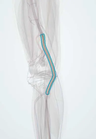
See it in 3D
Who gets it
Peripheral aneurysms are much less common than abdominal aortic aneurysms and mostly affect older men who smoke. Popliteal aneurysms are the commonest peripheral aneurysms, femoral aneurysms come next, and both are often bilateral (popliteal in about half to two thirds of patients). A large share of people with a popliteal aneurysm also have an abdominal aortic aneurysm, while only about 3 percent of people with an abdominal aortic aneurysm have a popliteal one. Visceral artery aneurysms are rare; the splenic artery accounts for about 60 percent of them and the hepatic artery for about 20 percent. Unlike other aneurysms, splenic artery aneurysms are about four times more frequent in women, especially after several pregnancies.
Causes
- Degenerative (atherosclerotic) wall weakening: most popliteal, femoral and iliac aneurysms
- Pseudoaneurysm after arterial puncture or catheterisation, surgery, trauma or pancreatitis (visceral arteries)
- Infection (mycotic aneurysm): endocarditis, injection drug use at the groin
- Pregnancy and multiparity, portal hypertension and liver transplantation (splenic artery)
- Connective tissue disorders (Marfan, Ehlers-Danlos) and vasculitis (polyarteritis nodosa, Behçet disease)
- Fibromuscular dysplasia (renal artery)
Risk factors
- Male sex, older age
- Smoking
- Hypertension and atherosclerosis
- An aneurysm elsewhere (aorta or the other leg)
- Pregnancy and portal hypertension (splenic)
- Recent arterial puncture, pancreatitis or abdominal trauma (pseudoaneurysm)
Pathophysiology
Wall weakens → the artery dilates → flow slows and swirls in the sac → laminated thrombus lines the wall. In the limbs: thrombus breaks off → repeated small emboli silently block the tibial arteries one by one → finally the sac itself thromboses → acute limb ischaemia with no outflow vessels left to bypass into, which is why limb loss is high. In the abdomen: wall tension rises with diameter (and with the high flow and hormonal softening of pregnancy for the splenic artery) → the wall gives way → rupture into the peritoneum or retroperitoneum, or into the bowel or bile ducts → haemorrhagic shock. Large aneurysms also compress neighbours: a popliteal aneurysm can press on the popliteal vein (swelling, thrombosis) or the tibial nerve.
Classification
- By site: popliteal, femoral, iliac (common or internal), visceral (splenic, hepatic, superior mesenteric, coeliac, pancreaticoduodenal), renal
- True aneurysm (all wall layers) vs pseudoaneurysm (contained leak, higher risk of rupture)
- Shape: fusiform (whole circumference) or saccular (a side pouch)
- Presentation: asymptomatic, symptomatic (pain, compression, embolism) or ruptured
Symptoms and signs
- Often none: found on imaging or as a pulsatile mass
- Popliteal: a wide, easily felt pulse behind the knee; calf claudication; blue toes from emboli; sudden pain, pallor, coldness and numbness of the leg when it thromboses
- Popliteal compression: calf swelling (vein) or pain and tingling (nerve)
- Femoral: a pulsatile groin lump, groin pain, claudication or acute ischaemia
- Iliac and visceral: usually silent until rupture; sudden abdominal, flank or back pain with collapse
- Renal: sometimes flank pain, blood in the urine or high blood pressure
Diagnosis
- Clinical
- Feel both popliteal fossae and both groins in every patient with an abdominal aortic aneurysm, and the abdomen in every patient with a peripheral aneurysm. A popliteal pulse that is unusually easy to feel suggests an aneurysm. Check foot pulses and look for signs of emboli. Ask about pregnancy plans in women with a splenic artery aneurysm, and about recent catheterisation, surgery or injection drug use.
- Tests
- Blood tests are for the setting: blood count, coagulation, crossmatch and kidney function before repair; blood cultures when infection is suspected; a pregnancy test in women of childbearing age.
- Imaging
- Duplex ultrasound is the first test for popliteal and femoral aneurysms: it measures the diameter and thrombus and is repeated on the other leg; the aorta is scanned too. CT angiography maps the inflow, outflow arteries and anatomy for repair and is the main test for iliac, visceral and renal aneurysms, most of which are found incidentally on CT or ultrasound. Catheter angiography is used when treatment is endovascular.
Differential diagnosis
- Baker (popliteal) cyst, which also sits behind the knee but has no pulse and no flow on duplex
- Peripheral artery disease with acute thrombosis or embolism from the heart
- Deep vein thrombosis (calf swelling from venous compression can mimic it)
- Groin lymph node or hernia
- Other causes of abdominal pain and shock: ruptured abdominal aortic aneurysm, placental abruption, ectopic pregnancy, splenic injury
Peripheral and visceral aneurysms: how each one harms
| Site | Typical patient | Main danger | Look for | Principle of treatment |
|---|---|---|---|---|
| Popliteal | Older male smoker | Thrombosis and emboli to the leg; rupture is rare | The other knee (often bilateral) and the aorta | Repair when symptomatic or at guideline size or thrombus; for acute ischaemia, clot removal plus bypass or covered stent |
| Femoral | Older male; after catheterisation (pseudoaneurysm) | Thrombosis, ischaemia, sometimes rupture | Aorto-iliac aneurysms (very common) | Repair large or symptomatic ones; puncture pseudoaneurysms are often treated with guided techniques |
| Iliac | Older male, often with an aortic aneurysm | Rupture, deep in the pelvis and silent | The aorta | Endovascular or open repair, usually with the aortic aneurysm |
| Splenic | Woman after several pregnancies; portal hypertension | Rupture, above all in pregnancy | Pregnancy plans; liver disease | Coil embolisation or covered stent; treat any size in pregnancy or when pregnancy is possible |
| Renal | Hypertension, fibromuscular dysplasia | Rupture (rare); renal infarct | Blood pressure, kidney function | Stent-assisted coiling or surgery, protecting the kidney's branches |
Goal of treatment
Prevent the specific catastrophe each site threatens (limb loss behind the knee, rupture in the abdomen), screen for aneurysms elsewhere, and choose open or endovascular repair according to anatomy, urgency and the patient's fitness.
Treatment
- Follow-up
- Small asymptomatic aneurysms: surveillance with duplex ultrasound or CT at intervals set by the guideline, plus control of risk factors (stopping smoking, blood pressure, lipids).
- Surgical
- Popliteal aneurysm: elective repair when symptomatic, or when size or thrombus reaches the guideline threshold, by excluding the sac and bypassing it with a vein graft, or by a covered stent graft in selected anatomy; stents behind the knee can kink or block with knee flexion.
- Acute
- Thrombosed popliteal aneurysm with acute limb ischaemia: anticoagulation and urgent revascularisation; catheter-directed thrombolysis or mechanical thrombectomy reopens the outflow arteries before or during open bypass or stent grafting and improves patency and limb salvage. Fasciotomy when compartment syndrome threatens.
- Procedure
- Visceral and renal aneurysms: endovascular coil embolisation, stent-assisted coiling or a covered stent in most stable patients; the spleen survives occlusion of the splenic artery through collateral short gastric arteries. Pseudoaneurysms, mycotic aneurysms and pancreaticoduodenal aneurysms are treated whatever their size.
- Acute
- Ruptured iliac or visceral aneurysm: resuscitation with a massive transfusion protocol and emergency endovascular control or surgery; in pregnancy a joint obstetric and surgical team.
Complications
- Acute limb ischaemia and amputation (popliteal, femoral)
- Distal embolism with loss of the tibial outflow arteries
- Rupture with haemorrhagic shock (iliac, visceral, renal)
- Rupture into bowel or bile ducts with gastrointestinal bleeding
- Compression of nearby veins or nerves
- After repair: graft thrombosis or kinking, endoleak, renal or splenic infarction
Common traps
- The danger of a popliteal aneurysm is rupture: it rarely bursts; it clots or throws emboli and costs the leg, so it is repaired to protect the limb
- A popliteal aneurysm is a local problem of one knee: it is often bilateral and travels with abdominal aortic aneurysms; scan the other knee and the aorta
- A pulsatile mass behind the knee is a Baker cyst: a cyst has no pulse and no flow; duplex ultrasound tells them apart before anyone puts a needle in it
- Blue toes with good foot pulses rule out an arterial cause: small emboli from a popliteal or femoral aneurysm block toe arteries while the main foot pulses remain
- A small splenic artery aneurysm can always be watched: in pregnancy, or when pregnancy is possible, any size is treated, because rupture is often fatal for mother and fetus
Prognosis
Elective repair of a popliteal aneurysm has good results; once it has thrombosed with acute ischaemia, amputation rates are high, which is why aneurysms are repaired before that happens. Visceral and renal aneurysms treated electively, mostly by endovascular means, do well, but rupture carries high mortality, especially for splenic aneurysms in pregnancy and for pancreaticoduodenal aneurysms.
Prevention
- Stopping smoking and controlling blood pressure
- Examining the popliteal fossae and groins in every patient with an abdominal aortic aneurysm, and scanning the aorta in every patient with a peripheral aneurysm
- Screening and repairing splenic artery aneurysms in women planning pregnancy
- Careful arterial puncture technique and compression after catheterisation
Test yourself
Think of the answer, then tap the question to see it.
A 72-year-old smoker has a sudden cold, numb leg and a firm pulseless mass behind the knee. What happened, and why is limb loss so likely?
A popliteal aneurysm has thrombosed. Earlier silent emboli have often blocked the tibial arteries, so there is little outflow left to restore; urgent thrombolysis or thrombectomy plus repair.
A man is found to have a popliteal aneurysm on one side. Which two other places do you image, and why?
The other popliteal artery, because about half are bilateral, and the abdominal aorta, because many coexist with an aortic aneurysm.
Why is a popliteal aneurysm more likely to thrombose, while a splenic artery aneurysm is more likely to rupture?
Behind the moving knee, slow, swirling flow lays down thrombus that blocks or embolises; in the abdomen the wall has no support, so rising tension, worse in pregnancy, ends in rupture.
Why can most splenic artery aneurysms be blocked with coils without removing the spleen?
The spleen has a collateral supply from the short gastric arteries, so it survives occlusion of the splenic artery.
Connections
Educational information, not medical advice. For any symptom, see a health professional.
Sources: Emergency endovascular management of peripheral artery aneurysms and pseudoaneurysms – a review (World Journal of Emergency Surgery (PMC), 2008); Comparisons of outcomes of open surgery versus endovascular intervention for thrombotic popliteal artery aneurysm with acute lower limb ischemia: a systematic review (BMC Surgery (PMC), 2022); Current management strategies for visceral artery aneurysms: an overview (Surgery Today (PMC), 2020); CIRSE Standards of Practice for the Endovascular Treatment of Visceral and Renal Artery Aneurysms and Pseudoaneurysms (CardioVascular and Interventional Radiology (PMC), 2024).
Spotted a mistake?We are always improving for the community. Tell us what is wrong and our team will review it.Report a mistake
Representative model, for educational purposes only.